Has distinct features of both epidermoid and pilar cyst
Diagnostic Checklist
• Abrupt tricholemmal keratinization is key diagnostic feature
• Simple unilocular cyst
If architectural complexity is present, consider proliferating pilar cyst or malignant pilar cyst
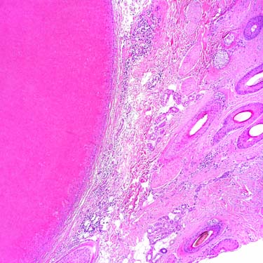
Pilar Cyst Involving Scalp The scalp is the most common location for pilar cysts, though they can rarely occur almost anywhere.
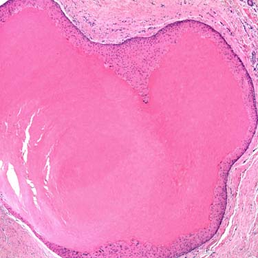
Well-Circumscribed Simple Cyst Pilar cysts are well-circumscribed simple cysts involving the dermis &/or subcutis. No granular layer is seen, even at low magnification.
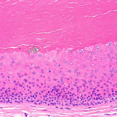
High-Power Image of Pilar Cyst The epithelium at the interface with the keratin is composed of large polygonal keratinocytes with abundant eosinophilic cytoplasm. There is abrupt keratinization without a granular layer, but scattered keratohyaline granules may be seen.
Ruptured Pilar Cyst Pilar cysts often rupture, resulting in granulomatous inflammation. Recognition of cyst remnants help make the diagnosis.
TERMINOLOGY
Synonyms
• Pilar cyst
• Trichilemmal cyst
• Isthmus-catagen cyst
Definitions
• Benign unilocular cyst lined by squamous epithelium lacking granular layer and containing abundant dense keratin material
CLINICAL ISSUES
Epidemiology
• Incidence
2nd most common type of cutaneous cyst
Only gold members can continue reading. Log In or Register to continue


 without a granular layer, but scattered keratohyaline granules may be seen.
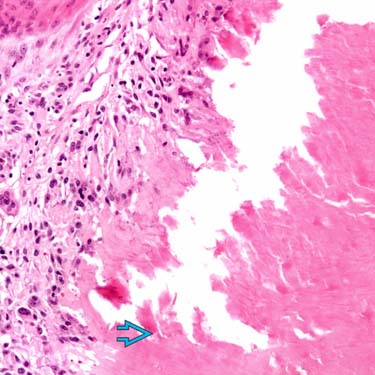
 help make the diagnosis.


