Patient Story
An African American medical student presented with a new dark band on her index finger for 1 year (Figure 191-1). The dark color and the lack of melanonychia in other fingers made this concerning. A biopsy of the nail matrix was performed and the result showed a benign nevus.
Figure 191-1
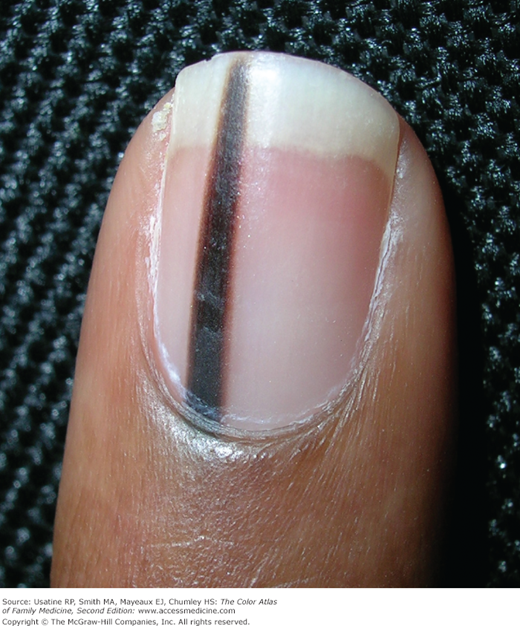
Longitudinal melanonychia—a single dark band of nail pigment appearing in the matrix region and extended to the tip of the nail. This is concerning for melanoma. The widening of the band in the proximal nail shows that the melanocytic lesion in the matrix is growing. This young woman had a biopsy that showed a benign nevus. (Courtesy of Richard P. Usatine, MD.)
Introduction
Atypical pigmentation of the nail plate may result from many nonmalignant causes, such as longitudinal melanonychia (LM), inflammatory changes, benign melanocytic hyperplasia, nevi, drugs, and endocrine disorders. It may also result from development of subungual melanoma. The challenge for the clinician is separating the malignant from the nonmalignant sources.
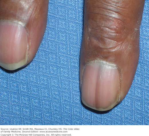
LM is the most common cause and it represents a longitudinal pigmented band in the nail plate (Figures 191-1 and 191-2). It may involve of 1 or several digits, vary in color from light brown to black, vary in width (most range from 2 to 4 mm), and have sharp or blurred borders.
Epidemiology
- LM is more common in more darkly pigmented persons. It occurs in 77% of African Americans older than age 20 years and in almost 100% of those older than age 50 years.1,2 It also occurs in 10% to 20% of persons of Japanese descent. LM is common in Hispanic and other dark-skinned groups. LM is unusual in whites, occurring in only approximately 1% of the population.1
- Melanoma is the seventh most common cause of cancer in patients in the United States. Subungual melanoma is a relatively rare tumor with reported incidences between 0.7% and 3.5% of all melanoma cases in the general population.3
Etiology and Pathophysiology
- LM originates in the nail matrix and results from increased deposition of melanin within the nail plate. This deposition may result from greater melanin synthesis or from an increase in the total number of melanocytes. Pigment clinically localized within the dorsal half of the nail plate indicates a proximal matrix origin, and pigment localized within the ventral nail plate indicates a distal matrix origin. Look at the distal edge of the nail in a cross-sectional view to see whether the pigment is dorsal or ventral (a dermatoscope may help).
- LM may also be caused by chronic trauma, especially in the great toes.
- Inflammatory changes accompanying skin diseases located in the nail unit, such as psoriasis, lichen planus, amyloidosis, and localized scleroderma, rarely may result in LM.
- Benign melanocytic hyperplasia (lentigo) is observed in 9% of the adult cases and 30% of the pediatric cases of single-biopsied LM.4
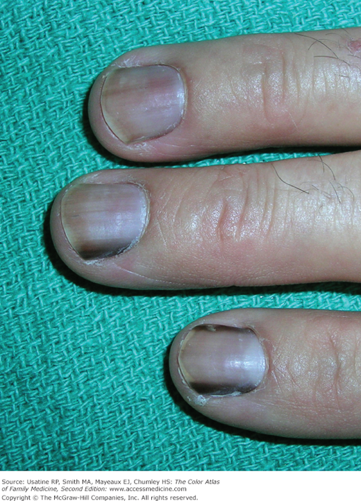
- Nevi represent 12% of LM in adults, but almost 50% of cases in children. A brown-black coloration is observed in two thirds of the cases and periungual pigmentation (benign pseudo-Hutchinson sign) in one third.
- Certain drugs may also cause LM, especially chemotherapeutic agents (Figure 191-3), and antimalarial drugs (mepacrine, amodiaquine, and chloroquine).
- Endocrine disorders, such as Addison disease, Cushing syndrome, hyperthyroidism, and acromegaly, can be responsible for LM.
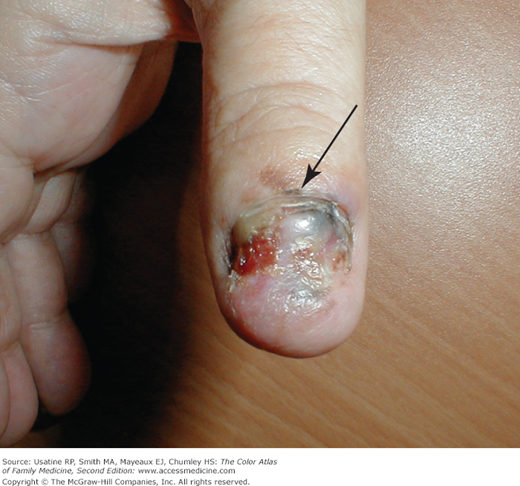
- The diagnosis of subungual melanoma must always be considered in patients with LM (Figures 191-4 and 191-5). Separating benign from malignant lesions is often difficult. Both arise most often in the thumb or index fingers, and both are more common in dark-skinned persons.5 A biopsy should be performed in an adult if the cause of LM is uncertain. Table 191-1 lists diagnostic clues for subungual melanomas.
- Hutchinson sign is the extension of pigmentation to the skin adjacent to the nail plate involving the nail folds or the fingertip. It is an important indicator for nail melanoma (Figures 191-4, 191-5, and 191-6).6
- Pseudo-Hutchinson sign is the presence of dark pigment around the proximal nail fold secondary to benign conditions such as racial melanosis and not melanoma (Figure 191-7). Another cause of pseudo-Hutchinson sign is a translucent cuticle below which the pigment of LM is visible. Trauma and drug-induced pigmentation can also produce a pseudo-Hutchinson sign.
- Subungual melanoma arises on the hand in 45% to 60% of cases, and most of those occur in the thumb (Figures 191-4, 191-5, and 191-6).4 On the foot, subungual melanoma usually occurs in the great toe.5 The median age at which subungual melanoma is usually diagnosed is in the sixth and seventh decades. It appears with equal frequency in males and females.5