Pigmented Epithelioid Melanocytoma (“Animal-type Melanoma”)
Soheil Sam Dadras, MD, PhD
Olubukola Babalola
Key Facts
Clinical Issues
Heavily pigmented, dome-shaped nodule
Occur sporadically or in association with Carney complex
Low-grade malignant potential
Complete excision necessary
Microscopic Pathology
Deep dermal tumor with frequent involvement of subcutis
More cellular in center and shows infiltrative growth pattern at periphery
Composed of 3 principal cell types
Medium-sized epithelioid cells, large epithelioid cells, and spindled cells
Top Differential Diagnoses
Malignant blue nevus
Atypical cellular blue nevus
Cellular blue nevus
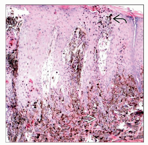 A punch biopsy specimen from an 11-year-old girl shows a heavily pigmented compound melanocytic tumor with epidermal hyperplasia, intraepidermal melanophages  , and large epithelioid cells , and large epithelioid cells  . . |
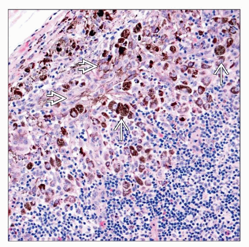 A sentinel lymph node biopsy specimen from an 11-yearold girl with PEM shows melanophages  and large epithelioid tumor cells and large epithelioid tumor cells  . These cells colonized multiple foci throughout the lymph node. . These cells colonized multiple foci throughout the lymph node. |


