Perineurioma
Thomas Mentzel, MD
Key Facts
Terminology
Benign mesenchymal neoplasm composed of neoplastic perineural cells
Clinical Issues
Progressive muscle weakness &/or sensory disturbances are seen in intraneural perineurioma
Extraneural perineurioma is not associated with neurofibromatosis
Macroscopic Features
Intraneural perineurioma is characterized by fusiform swelling of affected nerves
Extraneural perineuriomas are solitary, well-circumscribed, unencapsulated, nodular neoplasms
Microscopic Pathology
Residual S100 protein(+) nerve fibers are surrounded by EMA(+) perineural tumor cells in intraneural perineurioma
Different growth patterns (storiform, lamellar, fascicular) are present in extraneural perineurioma
Spindled tumor cells with elongated spindled nuclei and long and very thin cell processes
In addition, round, slightly enlarged tumor cells are seen
Sclerosing perineurioma is composed of plump spindled and epithelioid tumor cells
Prominent degenerative myxoid &/or edematous stromal changes are present in reticular perineurioma
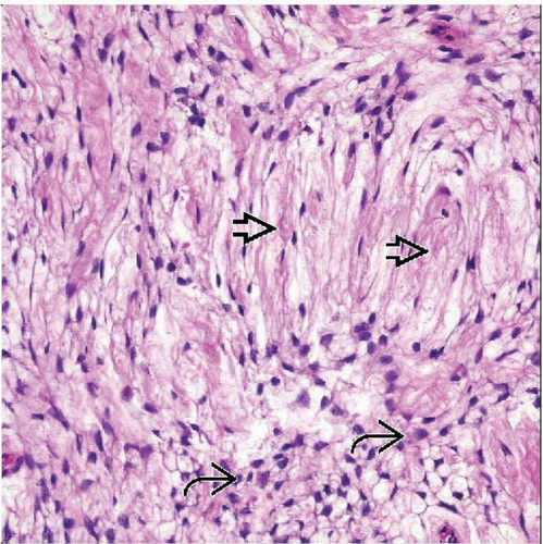 Perineurioma is composed of spindled tumor cells containing uniform fusiform nuclei and thin, elongated cell processes  . In addition, plump tumor cells with round nuclei are seen . In addition, plump tumor cells with round nuclei are seen  . . |
 Immunohistochemical staining for perineural markers (e.g., claudin-1) highlights the presence of thin and elongated cell processes in neoplastic perineural cells. |
TERMINOLOGY
Synonyms
Intraneural perineurioma (localized hypertrophic neuropathy of the limbs)
Extraneural perineurioma (storiform perineural fibroma)
Sclerosing perineurioma
Definitions
Benign mesenchymal neoplasm composed of neoplastic perineural cells
CLINICAL ISSUES
Epidemiology
Incidence
Very rare neoplasms
Age
Intraneural perineuriomas occur in adolescents and in early adulthood
Extraneural perineuriomas occur in adults of all ages
Children are only rarely affected
Gender
Intraneural perineurioma shows no sex predilection
Extraneural perineurioma shows slight female predominance
Site
Intraneural perineurioma is seen in peripheral nerves of limbs
Extraneural perineurioma arises most frequently on trunk and extremities
Sclerosing perineurioma tends to occur in superficial tissue of hands
Natural History
Progressive muscle weakness &/or sensory disturbances are seen in intraneural perineurioma
Extraneural perineurioma is not associated with neurofibromatosis
Treatment
Surgical approaches
Resection of affected nerves should be avoided as long as possible in intraneural perineurioma
Complete excision is advised in extraneural perineurioma
Prognosis
Intraneural perineuriomas are benign mesenchymal neoplasms
Malignant peripheral nerve sheath tumors with perineural differentiation (malignant perineuriomas) are extremely rare
MACROSCOPIC FEATURES
General Features
Intraneural perineurioma
Characterized by fusiform swelling of affected nerves
Segmental enlargement of affected nerve
Extraneural perineurioma
Solitary, well-circumscribed, unencapsulated, nodular neoplasms
Frequently in subcutaneous tissue, whereas deep soft tissue and dermis are more rarely affected
Sclerosing perineurioma
Arises more frequently in superficial dermal location
MICROSCOPIC PATHOLOGY
Histologic Features
Intraneural perineurioma
Residual S100 protein(+) nerve fibers are surrounded by EMA(+) perineural tumor cells
EMA(+) perineural cells form concentric layers around nerve fibers with characteristic pseudo-onion bulbs
Extraneural spindle cell perineurioma
Variable cellularity
Different growth patterns (storiform, lamellar, fascicular)
Spindled tumor cells with elongated spindled nuclei
Round, slightly enlarged tumor cells
Collagenous stroma may show focal hyalinization
Presence of focal infiltration and cytologic atypia does not affect benign biologic behavior
Rare hybrid forms of perineurioma/schwannoma and perineurioma/neurofibroma have been reported
Sclerosing perineurioma
Composed of plump spindled and epithelioid tumor cells
Hyalinized stroma containing numerous thin-walled blood vessels
Perivascular and lace-like arrangement of neoplastic cells often seen
Neoplasms showing combination of extraneural spindle cell and sclerosing perineurioma have been reported
Reticular perineurioma
Prominent degenerative myxoid &/or edematous stromal changes
Pseudocystic spaces may be present
Tumor cells with thin and elongated cell processes anastomose in lace-like, reticular pattern
Occasionally degenerative cytologic atypia is noted
Plexiform perineurioma
Extremely rare morphologic variant
Plexiform architecture of neoplastic perineural cells
Sclerosing pacinian-like perineurioma, lipomatous perineurioma, ossifying perineurioma, and perineurioma with granular cells represent very rare morphologic variants
Stay updated, free articles. Join our Telegram channel

Full access? Get Clinical Tree


