Some commonly used dermatological terms
Macule – a flat skin lesion <5 mm in diameter
Patch – a flat skin lesion >5 mm in diameter
Papule – a raised skin lesion <5 mm in diameter
Plaque – a raised skin lesion >5 mm in diameter
Nodule – a solid raised skin lesion that extends into the dermis or subcutaneous tissue
Erosion – partial loss of the epidermis
Ulcer – full-thickness loss of the epidermis
Vesicle – a fluid-filled blister <5 mm in diameter
Bulla – a fluid-filled blister >5 mm in diameter
Pustule – a pus-filled blister
Xerosis– dry skin
Telangiectasia – permanent dilation of small blood vessels in the skin
Koebnerization – appearance of rash/lesions of the same morphology at sites of trauma
Lichenification – thickening of the skin due to repeated rubbing
History
With a careful dermatological history (Box 20.1) and where relevant, a sexual history (Box 20.2), a differential diagnosis can often be deduced with clinical assessment alone (Table 20.2).
Table 20.2
Differential diagnoses
The multifactorial nature of perianal complaints can combine to produce a large differential diagnosis for any particular symptom. This box is a guide to possible diagnoses to consider |
White patches |
Vitiligo |
Lichen sclerosus |
AIN (see premalignant conditions) |
Lichen simplex |
Red lesions |
Dermatitis/eczema |
Atopic |
Contact |
Irritant/chemical |
Allergic |
Seborrhoeic |
Psoriasis |
Infection – bacterial or fungal |
Vascular tumours, e.g. hemangioma |
Fissures of the skin |
Crohn’s (multiple, ± anal canal fissure) |
Psoriasis |
Dermatitis |
Herpes simplex virus (HSV) |
Itching – see Pruritus Ani Chap. 21 |
Ulcer |
Herpes (HSV) |
Syphilis |
Lymphogranuloma venereum (LGV) |
Human immunodeficiency virus (HIV) |
Malignancy (see Malignant Disease) |
Crohn’s disease |
Varicella zoster |
Cytomegalovirus (CMV) |
Nicorandil |
Behçet’s disease |
Trauma/factitious |
Deep mycoses |
Chancroid |
Donovanosis (granuloma inguinale) |
Blistering disorders |
Vesicles |
Varicella including varicella-zoster virus |
Herpes simplex |
Stevens-Johnson syndrome |
Bullae |
Pemphigus vulgaris |
Bullous drug eruption, e.g. fixed drug eruption |
Lumps |
Lipoma |
Epidermoid cyst |
Dermoid cyst/teratoma |
Warts (condylomata acuminata) |
Molluscum contagiosum |
Condylomata lata |
Seborrhoeic keratoses |
AIN/bowenoid papulosis |
Pseudofolliculitis (ingrowing hairs) |
Prominent skin folds |
Box 20.1 Dermatological History
Presenting symptoms |
Itching – especially at night |
Bleeding |
Pain/soreness (e.g. on defecation, constant/intermittent, generalized/localized sharp/dull) |
Lumps (e.g. warts) |
Concerns of patients re symptoms (especially anxiety and embarrassment) |
Past medical history, for example, Crohn’s disease, atopy and HIV |
Family history of skin disease, for example, psoriasis and atopic dermatitis |
Drug history including topical medication |
General habits including use of soaps, shower gel, baby wipes and deodorants |
Box 20.2 Tips on Taking a Sexual History
Be relaxed and ask questions as you would normally | |
Be non-judgmental | |
Prepare: | |
Request that accompanying persons leave for a moment | |
Explain to the patient that you need to ask some personal questions about their sex life as it may be related to their symptoms | |
Reassure them that the information will be kept confidential | |
Obtain permission to proceed | |
Suggested questions: | Why ask this? |
1. When was the last time you had sex? | Find out if sexually active and open conversation |
(a) Was that a regular partner or a casual partner? | Gives an idea about risk taking |
(b) Was the partner male or female? | Ascertain sexual orientation; risk of STIs |
(c) Is the patient or the partner from another country? | Risk assessment for STIs |
(d) With a condom? | Risk taking behaviour |
2. When did you last have sex with a different person | Rate of partner change is an important risk factor for STIs |
Clarify details as above | |
3. Have they ever had anal sex? | Important risk factor for perianal STIs |
If so, was a condom used? | |
4. For men: have you ever had sex with a man? | This may affect risk of HIV, hepatitis B, even if long ago |
5. Have you ever injected drugs or had a blood transfusion (especially in a developing country or many years ago)? | Risk for blood-borne viruses |
In all sexually active or intravenous drug-using (past or present) persons, a blood-borne virus (HIV, hepatitis B and C) and syphilis screen should be offered. Ask if they have been previously tested and if so when and what the results were | |
Examination
After inspection of the genital and perianal areas, digital and speculum examination of the vagina and sigmoidoscopy of the rectum should be considered. Examination of the scalp, oral cavity, hair and nails and wider cutaneous examination can then be targeted according to the findings, for example, the extensor surfaces of limbs in psoriasis and oral cavity for lesions in secondary syphilis.
Diagnosis
The perianal skin is an accessible site for tissue sampling if a diagnosis has not been arrived at using clinical assessment. Skin scrapings (for microscopy and culture), swabs and biopsy are the mainstays of investigation.
For the proctologist, it is important to consider viral swabs for herpes simplex virus as these are often not part of the normal surgical management (see later in this chapter).
Treatment
Simple treatments, such as the use of emollients as soap substitutes, moisturizers and barrier creams to protect the skin are highly effective general measures in the management of many skin conditions. These agents are particularly useful in the management of dry skin, pruritus, psoriasis and irritant and atopic dermatitis (eczema).
Local anaesthetics have a role in painful disorders such as fissure in ano and first episode of herpes simplex virus infection (HSV) and are generally well tolerated, although contact dermatitis has been described [3].
Antihistamines are used orally for pruritic lesions.
Antimicrobial agents have an important role. Many of these agents possess anti-inflammatory activity and are used to treat both infectious and non-infectious skin disease, for example, the use of tetracycline in hidradenitis suppurativa.
Antifungals are used in managing dermatophytosis (ring worm), candidiasis (thrush), subcutaneous and systemic fungal infections and in controlling malassezia yeasts in seborrhoeic dermatitis.
Topical corticosteroids are the mainstay of the management of allergic and inflammatory dermatological conditions. There are low, intermediate and high potency topical steroid preparations. Weak preparations can be bought over the counter (Box 20.3).
Chronic use of potent topical corticosteroids can lead to atrophy and telangiectasia. Therefore, it is preferable that long-term management of patients requiring such agents be carried out under medical supervision. With purely perianal use, topical steroids result in insignificant systemic absorption.
Steroid-sparing agents include calcineurin inhibitors (e.g. pimecrolimus, tacrolimus). They should be used with caution and with specialist medical advice due to concerns regarding possible carcinogenic effects with long-term use.
Tumour necrosis factor (TNF) alpha inhibitor drugs (e.g. infliximab, adalimumab) are effective in Crohn’s disease [4], and there is limited evidence for their use in hidradenitis suppurativa [5].
Other treatment options include imiquimod, retinoids and laser. Surgical management is necessary for abscesses, biopsy and excision.
Box 20.3 Topical Corticosteroid Preparation Potencies
Mild: |
Hydrocortisone 0.5 %; 1 % |
With antifungals: |
Hydrocortisone 1 % + clotrimazole 1 % |
Hydrocortisone 1 % + miconazole 1 % |
With antibacterials: |
Hydrocortisone 1 % + fusidic acid 2 % |
Moderate: |
Clobetasone butyrate 0.05 % |
With antimicrobials: |
Clobetasone butyrate 0.05 % + oxytetracycline 3 % + nystatin 10,000 units/g |
Potent: |
Betamethasone valerate 0.1 % |
Mometasone furoate 0.1 % |
Hydrocortisone butyrate 0.1 % |
With antimicrobials: |
Betamethasone valerate 0.1 % + fusidic acid 2 % |
Very potent: |
Clobetasol propionate 0.05 % |
Diflucortolone valerate 0.3 % |
Topical antibacterial preparation: |
Fusidic acid 2 % |
Mupirocin 2 % |
Topical antifungal preparation: |
Clotrimazole 1 % |
Miconazole 2 % |
Terbinafine 1 % |
Sexual Health for the Proctologist
Sexually transmitted infections (STIs) frequently involve the perianal area. Sexual practices involving the anus are common: anal intercourse in the last year was reported by more than 10 % of UK adults and more than 20 % of US adults in household surveys [6, 7]. Infections are discussed individually, but the following principles apply to management of all STIs:
2.
Confirm the diagnosis in the laboratory if at all possible. Ensure clinics and theatres are supplied with the equipment for viral and Chlamydia (nucleic acid amplification tests – NAATs) testing.
3.
Treat diagnosed infections quickly (e.g. herpes simplex infection) or refer rapidly for urgent treatment (e.g. for syphilis), recommending sexual abstinence in the meantime. Ensure local guidelines are followed.
4.
STIs travel in packs – always screen for other infections or recommend attendance elsewhere for screening, including HIV, if you suspect or diagnose one STI.
5.
Management of STIs is not complete without partner screening and treatment. Refer for management of partners or at least ensure the patient is aware that their partners are at risk.
6.
STI diagnoses are associated with stigma and psychological morbidity. Some patients may require referral for specialist support.
Non-infectious Skin Disorders
Dermatitis (Eczema)
Dermatitis is a general term to describe inflammation of the skin. It is often used synonymously with eczema, a term that derives from the Greek ‘to boil’. It describes a group of diseases which present with an itchy red rash that may show oozing, crusting or scaling.
Atopic Dermatitis (Atopic Eczema)
Atopic dermatitis/eczema is a pruritic eruption that is recurrent, usually flexural in adults, symmetrical and associated with a predisposition to atopy (personal or family history of asthma, eczema or hay fever). It can present in infancy, childhood or adulthood. Perianal involvement can rarely occur in all three stages. The napkin area is usually spared in infancy but can be affected [6]. Atopic dermatitis accounts for 3–5 % of cases of pruritus ani in adults [8]. Although atopic dermatitis is common, isolated involvement of the perianal area is rare.
Treatment involves the use of antihistamines to control itching and mild to moderately potent topical corticosteroid preparations to control inflammation.
Contact Dermatitis
Contact dermatitis is a common underlying or contributory cause of perianal symptoms and presents as an erythematous lesion representing a reaction to a substance which has breached the skin barrier [9].
Two forms of contact dermatitis are described which can be difficult to distinguish clinically: irritant contact dermatitis, where the cells are directly damaged by the agent in question, and allergic contact dermatitis, where such a substance induces a delayed-type hypersensitivity reaction. Predisposing factors to this multifactorial condition include atopic dermatitis, genetic factors and environmental cofactors such as moisture and occlusion [10].
Irritant Contact Dermatitis
This is caused by either exposure to weak irritants (e.g. soap, shower gel) or contact with strong irritants such as acids and alkalis and is a nonimmune-mediated inflammatory skin reaction presenting in acute or chronic forms.
Symptoms include irritation and soreness or a burning sensation. The skin usually appears erythematous with vesicles in the acute stage with scaling, fissures and lichenification in the chronic stage. Potential irritants in the perianal area include hygiene products, wet wipes, detergents, topical haemorrhoid preparations and products for the relief of itching which can paradoxically contain irritants as preservatives. Mucus and faecal matter from incontinence, prolapse of haemorrhoids, or from the rectal mucosa lead directly to irritant contact dermatitis, as it does in infants (diaper/nappy rash).
Treatment involves withdrawal of the irritant or protection against it with barrier creams along with application of weak or moderately potent topical corticosteroid cream or ointment.
Allergic Contact Dermatitis
Allergic contact dermatitis can be difficult to distinguish from irritant contact dermatitis. It is due to a delayed hypersensitivity reaction (type IV) [11]. An analysis of 1,168 suspected anogenital contact dermatitis patients showed that allergy was responsible in 25 %, with the main culprits being the patient’s own products as well as lanolin and fragrances, and hence, these should be used for allergen patch testing which is now widely available [12]. Nail varnish allergy has been described as a cause of perianal rash [13].
A preservative used in the increasingly popular moist toilet tissues/baby wipes (methylchloroisothiazolinone/methylisothiazolinone, MCI/MI) is known to cause allergic contact dermatitis [14]. Ingested allergens such as nickel [9] or spices in food, particularly those related to balsam of Peru (cinnamon, cloves, nutmeg, vanilla), can lead to perianal dermatitis [15]. More commonly, however, allergic contact dermatitis is due to preparations intended to ameliorate anal symptoms: haemorrhoid creams, deodorants and local anaesthetics [16]. Treatment involves identifying and removing contact with the allergen to which the patient is sensitized and use of mild to moderately potent topical corticosteroid preparations to control inflammation.
Seborrhoeic Dermatitis (Seborrhoeic Eczema, Cradle Cap)
Other affected sites: scalp, eyebrows, retroauricular and genitocrural.
Seborrhoeic dermatitis is a chronic inflammatory condition which can affect the perianal area. It is relatively common (3–5 %) and more prevalent in men. It is characterized by pruritic erythematous areas with greasy yellowish fine scaling located in areas of high sebum production, such as the scalp, back, groins, chest and the genitocrural region. Scaling may be less obvious in the perianal area, and examination of other typical sites will give a clue to the diagnosis.
Seborrhoeic dermatitis is etiologically associated with the yeast Malassezia furfur (Pityrosporum ovale), and the two ages of greatest incidence – infantile (2 weeks to 12 months) and adolescent-adult – correspond to periods of increased sebum production. However, the relationship is not simple. Treatment involves controlling seborrhoea of the scalp with antifungal shampoos such as ketoconazole or zinc pyrithione at the same time as low to intermediate potency topical steroids in combination with antifungals for the perianal skin lesions. Severe or treatment-resistant seborrhoeic dermatitis may require oral antifungals and is an indicator condition for unrecognized human immunodeficiency virus (HIV) infection [17].
Secondary bacterial infection can occur, and the differential diagnosis includes flexural psoriasis and intertrigo.
Lichen Simplex Chronicus (LSC)
This is a chronic eczematous condition characterized by localized plaques due to repeated rubbing and scratching in response to pruritic stimuli. The skin, initially erythematous, becomes lichenified. This leads to further itching which results in habitual rubbing and more lichenification, the ‘itch-scratch cycle’. Any area that can be conveniently reached can be affected including the perianal area where lesions can be asymmetrical (see Fig. 20.1). Precipitating factors include heat, sweat, chemical irritation and ‘psychological’. Twenty-five percent of patients have a background of atopy [18, 19].
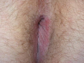
Fig. 20.1
Lichen simplex chronicus. Note lichenification over the perianal area especially on the left side (Courtesy of Dr P N Sashidharan, Homerton University Hospital, London)
LSC must be distinguished from psoriasis, contact dermatitis and anal intraepithelial neoplasia (AIN). All causes of pruritus ani need to be considered in the search for an underlying trigger, including candidiasis, dermatophyte infection, parasitic infestation as well as faecal incontinence, haemorrhoids and neoplasia (see Chap. 21). Topical steroids are the mainstay of treatment. Sedative antihistamines are helpful for controlling pruritus at night. Other measures include generic pruritus advice: avoiding soaps, using cotton anal plugs and barrier creams. Patients should be made aware that the rash will not resolve until scratching and rubbing are stopped.
Psoriasis (Inverse Psoriasis, Flexural Psoriasis)
Associated sites: extensor surfaces especially elbows, knees, scalp and nails.
Psoriasis is a chronic inflammatory disorder, which, in its most common form, is characterized by erythematous scaly papules and plaques typically seen over scalp and extensor surfaces of the body. These are generally asymptomatic but lesions occasionally can be pruritic.
Psoriasis affects 1–5 % of the population in Western societies, with bimodal peaks in the early 20s and mid-50s, with men and women equally affected. There are several types of psoriasis, but the type seen in the perianal area is the ‘flexural’ or ‘inverse’ type which also affects axillae, popliteal fossae and inguinal and intergluteal areas, as well as the inframammary area in women.
Well-defined erythematous plaques or patches with fissuring are seen which differ from psoriasis of non-intertriginous areas due to minimal scaling (see Fig. 20.2). Genital and anal lesions usually occur concomitantly. Although isolated perianal psoriasis can rarely occur, other sites are often affected and should be examined. Nail changes include irregular pits, onycholysis (separation of nail from the nail bed), subungual hyperkeratosis (accumulation of keratin material underneath the nail) and translucent yellowish spots under the nail plate (oil drop sign).
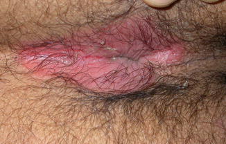
Fig. 20.2
Psoriasis. Well-defined erythematous plaque with fissuring. Scaling is usually not a feature at flexures. (Courtesy of Dr P N Sashidharan, Homerton University Hospital, London)
Perianal psoriasis can mimic dermatitis, fungal or bacterial infection due to the lack of characteristic scaling at this site.
Psoriasis may be the initial presentation of HIV infection, or HIV can aggravate existing psoriasis (see later in this chapter).
Treatment is with mild or moderately potent topical steroids with or without antifungal agents. Vitamin D analogues can be used as steroid-sparing agents.
Lichen Sclerosus (Lichen Sclerosus et Atrophicus and Balanitis Xerotic Obliterans, LS)
Additional site: vulva.
Lichen sclerosus is a dermatological disorder of unknown aetiology with predilection for the anogenital region. There is a peak of incidence in childhood. In adulthood, women are affected more than men (10:1), usually at a postmenopausal stage.
LS is considered an autoimmune disorder with a similar profile to vitiligo, morphea and autoimmune disease of the thyroid [20].
It is characterized by ivory-white papules that become confluent to form whitish plaques that subsequently become atrophic (see Fig. 20.3). Keratotic follicular plugs may be seen on the surface in early lesions. Focal areas of petechiae or purpura may be seen in established lesions.
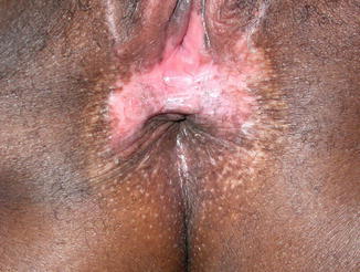
Fig. 20.3
Lichen sclerosus. Note whitish sclerotic area over perineum extending to the perianal area and hypopigmented macules and papules confined to the rest of the perianal area. The vulva was also affected (Courtesy of Dr P N Sashidharan, Homerton University Hospital, London)
In women, involvement of the vulva and perianal area simultaneously gives rise to a ‘figure of eight’ or ‘hour glass’ appearance. Scarring develops subsequently. Malignancy can occur in 3–4 % of cases of lichen sclerosus and needs to be excluded if plaques or ulcerated lesions develop.
Biopsy helps to confirm the diagnosis and exclude premalignant and malignant changes. The mainstay of treatment is potent topical steroid creams. Long-term follow-up is recommended in view of the potential for malignant change.
Lichen Planus (LP)
Additional sites: oral mucosa, trunk and nails.
Lichen planus is a chronic inflammatory pruritic mucocutaneous disease. The classic lesion is an erythematous flat-topped violaceous papule. Whitish reticular lines can be seen on the surface (Wickham’s striae). Koebner phenomenon can occur with new lesions developing at sites of injury. Whitish reticulated patches may be seen in the oral cavity, and nail changes include longitudinal ridging and pterygium (fusion of the nail fold to the matrix).
There are several types of LP. The papular and hypertrophic types can affect the natal cleft and buttocks.
The erosive type of LP affects the anogenital area and the oral cavity. It is painful due to the desquamation, is chronic and is recalcitrant to treatment. There is also a small (2–3 %) risk of developing squamous cell carcinoma.
Lichen planus usually responds to potent topical steroids. Tacrolimus or pimecrolimus have also been used with some success [21]. Refractory cases may require oral immunosuppression. Late presentation of erosive lichen planus can cause adhesions and lead to stenosis of the introitus or, rarely, of the anus.
Hidradenitis Suppurativa (Acne Inversa, Verneuil’s Disease, HS)
Associated sites: inguinal, axillae, inframammary and buttocks.
Hidradenitis suppurativa is defined as a ‘chronic, inflammatory, recurrent, debilitating skin disease’ [22]. It is a cause of chronic abscesses and sinus formation in apocrine gland-bearing areas of the skin – most commonly the axillae, groins, perianal and buttock areas.
HS is extremely rare before puberty and more common in women (3:1). Involvement of axillary, groin and inframammary areas is seen more in females, whereas the perianal region and buttocks are more affected in men. Around 25 % have a familial form with an autosomal dominant pattern [23].
The exact pathogenesis of HS is not known, but it is thought to be due to occlusion of the pilo-sebaceous-apocrine units which leads to rupture of the follicle and an aseptic chronic inflammatory process resulting in painful deep-seated red nodules and sinus formation [22].
There is an association with other follicular occlusion disorders leading to the concept of a tetrad which also includes pilonidal sinus, acne conglobata and dissecting cellulitis [24]. Other predisposing factors include smoking, obesity and altered sensitivity to hormones particularly androgens although their influence has yet to be fully elucidated [25].
The role of bacteria is debated: secondary infection may occur; a primary causal role is not established.
The clinical diagnostic hallmark is the presence of double or multiple comedones (‘tombstone’ comedones). The nodules do not point which distinguishes them clinically from bacterial furunculosis. The Hurley classification consists of three stages: stage one constitutes abscess formation and is the most common type (70 % of cases of HS); stage two involves sinus formation and cicatrisation (25 %); and 1 % progress to stage three severe disease with diffuse-bridged scarring and interconnecting tracts [26].
The differential diagnosis includes Crohn’s disease, furunculosis, infected epidermoid cysts and relatively rare infections such as tuberculosis, lymphogranuloma venereum, nocardia and actinomycosis.
Differential from cryptoglandular fistulation is usually made by inspection: HS is a superficial disease which although occasionally involving the anal canal does not fistulate across or within the sphincters [27] (see Fistula-in-Ano chapter). Despite this, the differential diagnosis can be difficult clinically and require both examination under anaesthetic and MRI scan of the perineum to exclude deep extension, as well as biopsy.
Diagnosis depends on the triad of features: typical clinical appearance, typical sites and the recurrent nature of the disease.
Conservative management includes general measures such as reducing heat, humidity and sweating with loose-fitting undergarments, local antiseptic washes, losing weight and stopping smoking.
Antibiotics are used and may be needed long term. The most effective are those with an anti-inflammatory function, such as the tetracyclines, clindamycin and rifampicin. Modulation of the sex hormones with anti-androgens is employed, for example, with the use of oral contraceptives containing cyproterone acetate. Retinoids also have been used therapeutically as have corticosteroids, zinc gluconate [28] and topical resorcinol treatment [29]. Nd: YAG laser therapy has found a place [30]. The role of TNF-alpha inhibitors is being explored: response in Hurley stage III can be rapid with a dramatic effect [31]. Surgery is used in three main ways: incision and drainage of abscesses, deroofing and wide excision. Surgery in combination with TNF-alpha inhibitors may be required for severe disease (see Fig. 20.4).
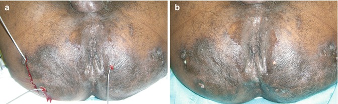
Fig. 20.4
Hidradenitis suppurativa: superficial fistulation and sinus tracts, (a) multiple fistulae of perianal hidradenitis suppurativa (b) probes in sinus tracks, perianal hidradenitis suppurativa (Courtesy of Mr P A Giordano, Whipps Cross University Hospital, London)
Crohn’s Disease (Crohn’s Disease, Terminal Ileitis)
Associated sites: ileum, colon, oral cavity and extra-intestinal.
It is recognised that the gastrointestinal tract from mouth to anus can be affected with the characteristic full-thickness non-caseating granulomatous inflammation of Crohn’s disease. There are extra-intestinal manifestations such as the rare cutaneous involvement with granulomatous inflammation (metastatic Crohn’s disease) as well as more commonly associated joint, eye, skin and hepatobiliary disorders.
Perianal Crohn’s disease represents a major part of Crohn’s morbidity, with pain and incontinence being common features. Perianal involvement in Crohn’s disease is fairly common, with 25 % of those with terminal ileal and 75 % of those with colonic Crohn’s affected, and the more distal the affected Crohn’s segment, the more likely the perianal involvement.
Crohn’s disease can affect ages from childhood onwards; however, it has a bimodal distribution with a peak at age 20–40 years and a second at ages 60–70 years. Crohn’s patients with perianal disease present 8 years younger than those without [32]; women are slightly more likely to have an associated perianal diagnosis (57 %) [33]; perianal disease at an early age is associated with a more severe course for both luminal and perianal Crohn’s [32, 34]. It is a risk factor for proctectomy and stoma formation [33, 35].
Perianal Crohn’s can predate the appearance of Crohn’s disease elsewhere which it behoves the clinician to consider [36] (see Fig. 20.5).
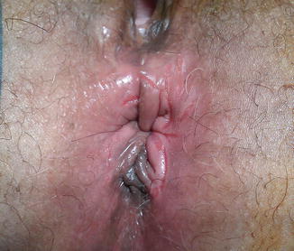
Fig. 20.5


Crohn’s disease of the perianal area without fistulation: skin tags and superficial fissuring (Courtesy of Mr P A Giordano, Whipps Cross University Hospital, London)
Stay updated, free articles. Join our Telegram channel
Full access? Get Clinical Tree
