Paraganglioma
Nina Gale, MD
Cyril Fisher, MD, DSc, FRCPath
Key Facts
Terminology
Tumor arising from paraganglia
Clinical Issues
Variety of sites
More frequent in head and neck
Thorax, abdomen, larynx, bladder
Functional or nonfunctional
Mostly benign
Microscopic Pathology
Circumscribed tumor
Nested alveolar (zellballen) pattern
Chief cells with granular cytoplasm
Clear cells in some
Nuclear pleomorphism not uncommon
Sustentacular cell processes around nests
Highly vascular stroma
Focal fibrosis
Malignant variants have mitoses, necrosis, vascular invasion, fewer sustentacular cells
Histology does not predict behavior
Ancillary Tests
CD56(+), chromogranin(+), neurofilament(+)
S100 protein(+) in sustentacular cells
Cytokeratin(-) and EMA(-)
Electron microscopy shows dense-core granules
Top Differential Diagnoses
Neuroendocrine carcinoma
Alveolar soft part sarcoma
Metastatic renal cell carcinoma
Adrenal cortical carcinoma
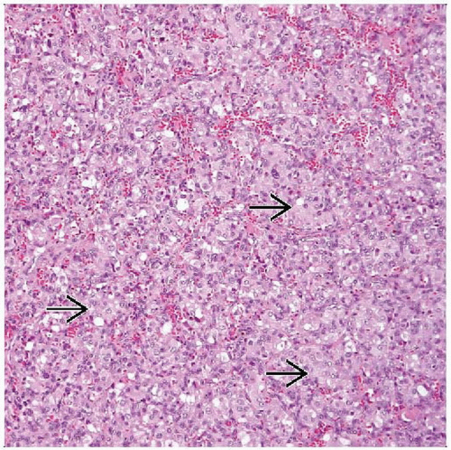 Hematoxylin & eosin shows the characteristic alveolar pattern (zellballen) of paraganglioma. Small nests of cells  are surrounded by a fibrovascular, richly vascularized stroma. are surrounded by a fibrovascular, richly vascularized stroma. |
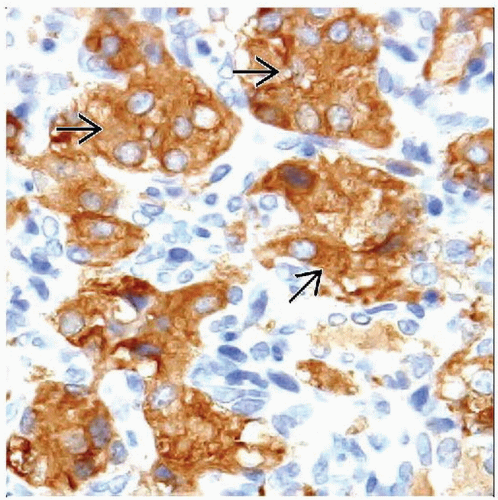 Synaptophysin shows strong and diffuse immunoreactivity of the chief cells  . There is no reactivity of the supporting sustentacular cells (which would stain with S100 protein). . There is no reactivity of the supporting sustentacular cells (which would stain with S100 protein). |
TERMINOLOGY
Synonyms
Chemodectoma
Glomus (do not confuse with soft tissue glomus)
Definitions
Neuroendocrine tumor arising from paraganglia at specified locations
Chromaffin(+) tumors resemble pheochromocytoma
Aorticosympathetic, can be functional
Chemoreceptor (branchiomeric) type are nonchromaffin
Usually nonfunctional
Chief and sustentacular cells arranged in organoid pattern
CLINICAL ISSUES
Epidemiology
Incidence
Sporadic or familial
Germline mutations include succinate dehydrogenase gene SDH
Can be part of MEN2, Carney triad, or NF1
Many familial lesions are multifocal
Age
Any age but mostly adults
Gender
M > F for carotid body tumors
Slight female predominance in other sites
Site
Head and neck
Carotid body is most common site
> 1/2 of head and neck paragangliomas
More common in high-altitude habitat
Jugular bulb, middle ear
Vagus nerve
Mediastinum (aorticopulmonary)
Retroperitoneum (from/aorticosympathetic paraganglia or organ of Zuckerkandl)
Organ-based
Duodenum (gangliocytic paraganglioma)
Bladder, heart, larynx
Presentation
Painless mass
Pressure effect
Vagal symptoms
Treatment
Surgical approaches
Surgical excision with external approach
Intraoperative bleeding may be significant
Prognosis
Most benign but can recur
Rare malignant examples
Metastasize to lymph nodes, lung, bone
MACROSCOPIC FEATURES
General Features
Circumscribed mass
Cut surface is pink to tan and dark red
Size
Range: 0.5-6 cm
MICROSCOPIC PATHOLOGY
Histologic Features
Nests of tumor cells in highly vascular fibrous tissue
Chief cells have eosinophilic, finely granular cytoplasm and central nuclei
Nuclear pleomorphism may be present but is prognostically unimportant
Clear cells change focally or diffusely
Sustentacular cells at periphery of nests
Modified Schwann cells
Rare mitoses
Sinusoidal vascular pattern
Malignant features include
Marked nuclear pleomorphism, mitotic activity
Necrosis, vascular invasion
Paucity of sustentacular cells
Occurrence of metastasis
Hyalinizing variant
ANCILLARY TESTS
Electron Microscopy
Chief cells have dense core granules 100-400 nm
Sustentacular cells have continuous external lamina
Stay updated, free articles. Join our Telegram channel

Full access? Get Clinical Tree


