Most cases occur in adults but some occur in children
May show significant overlap with Dabska tumor, with prominent papillary structures
• Composite hemangioendothelioma
Composed of at least 2 distinct HE types
• Kaposi sarcoma
Proliferation of small, slit-like vascular spaces lined by HHV8(+) spindle cells
Lacks papillary pattern of Dabska
• Angiosarcoma
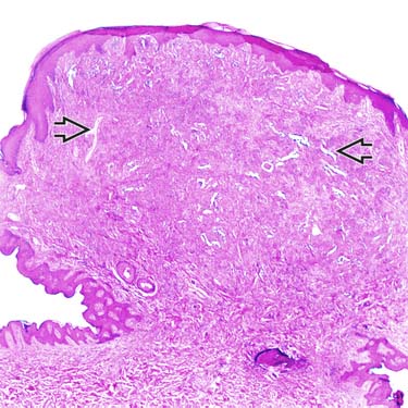
Scanning Magnification of Dabska Tumor Scanning magnification view of a papillary intralymphatic angioendothelioma (Dabska tumor) shows a polypoid lesion in the dermis with irregularly dilated vascular spaces .
Low Magnification of Dabska Tumor Low magnification shows a background of lymphangioma-like areas with scattered lymphoid aggregates . Note the focal intraluminal papillary projections .
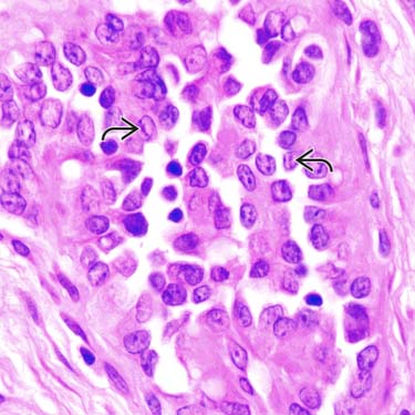
High Magnification Showing Cytologic Details High power shows papillae lined by mildly enlarged hobnailed cells projecting into the vascular lumina. The cells show vesicular chromatin, small nucleoli, and occasional grooves .
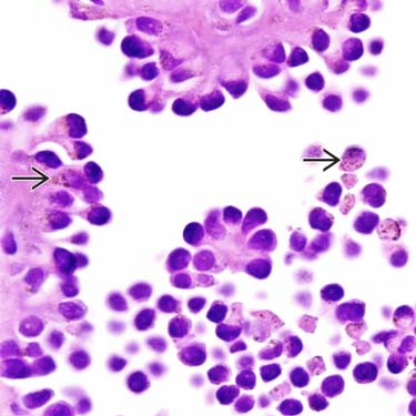
Papillae Lined by Plump Endothelial Cells Multiple prominent intralymphatic projections of papillary structures with hyaline cores are lined by plump, hyperchromatic-staining endothelial cells, some of which contain cytoplasmic hemosiderin pigment .
TERMINOLOGY
Synonyms
• Dabska tumor
• Endovascular papillary angioendothelioma
Definitions
• Low-grade malignant vascular tumor composed of hobnailed endothelial cells
ETIOLOGY/PATHOGENESIS
Unknown
• May be associated with vascular or lymphatic tumor/malformation
CLINICAL ISSUES
Epidemiology
• Incidence
Rare tumors
• Age
Typically occur in children (minority in adults)
• Sex
Slight female predominance
Site
• Distal extremities most common but may occur in other sites
Only gold members can continue reading. Log In or Register to continue


 .
.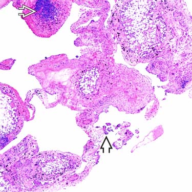
 . Note the focal intraluminal papillary projections
  .
.
 .
.
 .
.


