PEH lacks nuclear atypia, tumor cell necrosis, and mitotic activity present in angiosarcoma
• Hemangioma
• PEH-like changes may occur in hemangiomas complicated by thrombosis
• Arteriovenous malformation (AVM)
PEH-like changes may occur in background of AVM
• Hematoma
• Venous lymphatic malformation (VLM)
In VLM, there are lobules of abnormally dilated vessels, some with lymphatic morphology
Well-Circumscribed Vascular Proliferation Papillary endothelial hyperplasia (PEH) is a well-circumscribed reactive lesion in which papillary fronds lined by a single layer of endothelial cells proliferate within a vascular lumen .
Fibrin Thrombi Seen in PEH Fibrin thrombi are apparent in early stages, and with time are replaced by papillary fronds with a fibrous core lined by a single layer of banal-appearing endothelial cells characteristic of PEH.
PEH With Single Layer of Flat Endothelium The lined endothelium is a single layer of evenly spaced, banal-appearing flat cells wrapping around the fibrotic core .
Some Cases of PEH Can Resemble Angiosarcoma PEH can be mistaken for the dissecting vascular spaces seen in low-grade angiosarcoma. Note the bland endothelial lining of the blood vessel lumen and intravascular location.
TERMINOLOGY
Abbreviations
• Papillary endothelial hyperplasia (PEH)
Synonyms
• Masson tumor
• Vegetant intravascular hemangioendothelioma
• Intravascular angiomatosis
• Intravascular papillary endothelial hyperplasia
Definitions
• Benign, reactive, and papillary endothelial proliferation within vessel
ETIOLOGY/PATHOGENESIS
Reactive Vascular Proliferation
• Manifestation of organizing intravascular thrombus
• PEH-like changes may be present in preexisting hemangiomas or vascular malformations
CLINICAL ISSUES
Site
• Wide distribution
• Common sites include: Head and neck, fingers, trunk
Presentation
• Painless mass
• Located in deep dermis &/or subcutaneous tissue
Treatment
• Excision is not necessary but is curative
Prognosis
• Excellent
• Cases with underlying hemangioma or vascular malformation may recur
MACROSCOPIC
General Features
• Cystic mass with red-purple discoloration
• Often surrounded by pseudocapsule
Size
• Mostly small (< 2 cm)
MICROSCOPIC
Histologic Features
• Circumscribed lesion with pseudocapsule
Residual smooth muscle or elastic lamina of preexisting vessel may be apparent
Only gold members can continue reading. Log In or Register to continue


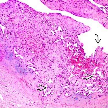
 lined by a single layer of endothelial cells proliferate within a vascular lumen
  .
.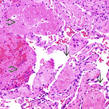
 are apparent in early stages, and with time are replaced by papillary fronds with a fibrous core lined by a single layer of banal-appearing endothelial cells
  characteristic of PEH.
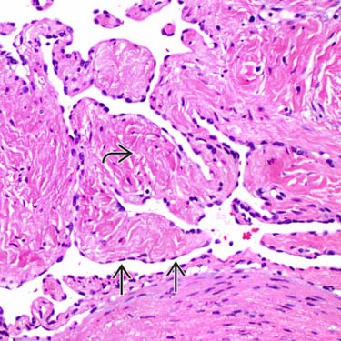
 is a single layer of evenly spaced, banal-appearing flat cells wrapping around the fibrotic core
  .
.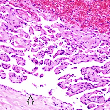
 of the blood vessel lumen and intravascular location.


