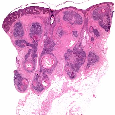
Low-Power Image of Panfolliculoma Panfolliculoma is characterized by a dermal-based adnexal proliferation recapitulating all levels of the hair follicle.
Medium-Power Image of Panfolliculoma Resembling Primitive Follicles Panfolliculoma shows cystic areas lined by stratified squamous epithelium with a granular layer , basaloid epithelium with papillary mesenchymal bodies , and inner and outer root sheath .
Higher Power Image of Panfolliculoma With Component Resembling Infundibulum The cystic areas are lined by stratified squamous epithelium with a granular layer and filled with abundant laminated keratin , consistent with infundibular differentiation.
High-Power Image of Panfolliculoma With Hair Bulb Formation There is advanced follicular differentiation with external and internal root sheath formation, as well as matrical epithelium. At the base there is a condensation of spindled cells representing a papillary mesenchymal body .
Only gold members can continue reading. Log In or Register to continue


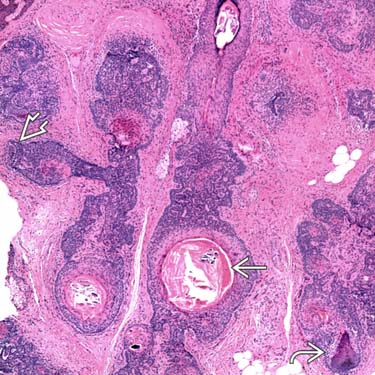
 , basaloid epithelium with papillary mesenchymal bodies
  , and inner and outer root sheath
  .
.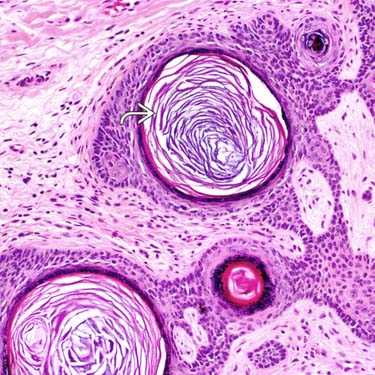
 , consistent with infundibular differentiation.
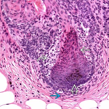
 and internal
  root sheath formation, as well as matrical
  epithelium. At the base there is a condensation of spindled cells representing a papillary mesenchymal body
  .
.


