Palisaded Myofibroblastoma
Cyril Fisher, MD, DSc, FRCPath
Key Facts
Terminology
Benign spindle cell tumor of modified smooth muscle cells in lymph node
Clinical Issues
Mainly inguinal region
Mostly in males
Macroscopic Features
Cut surface often dark red or black
Microscopic Pathology
Rim of lymph node tissue
Focal palisading, amianthoid fibers
Ancillary Tests
Actin(+), desmin(-), and CD34(-)
Top Differential Diagnoses
Kaposi sarcoma
Schwannoma
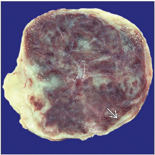 Gross pathology photograph shows a hemorrhagic tumor extensively replacing the lymph node. Residual nodal tissue is seen at the periphery  . . |
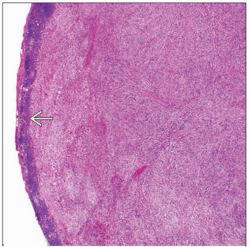 Hematoxylin & eosin shows a cellular tumor with irregular fascicles and vague palisades of uniform spindle cells. Note rim of compressed lymph node  . . |
TERMINOLOGY
Synonyms
Intranodal hemorrhagic spindle cell tumor with amianthoid fibers
Solitary spindle cell tumor with myoid differentiation of lymph node
Definitions
Benign spindle cell tumor of modified smooth muscle cells in lymph node
CLINICAL ISSUES
Epidemiology
Incidence
Rare
Mainly inguinal region
Occasionally in submandibular lymph node
Rarely multicentric
Age
5th and 6th decades
Gender
Stay updated, free articles. Join our Telegram channel

Full access? Get Clinical Tree


