Patient Story
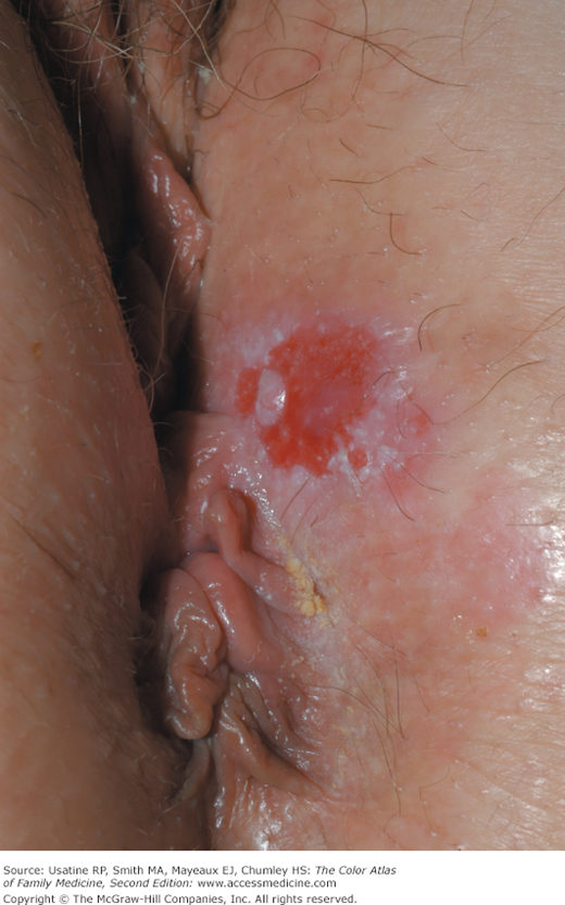
A 60-year-old woman presented with vulvar pruritus for 1 year that is now constant. On physical examination, there is one large red lesion surrounded by white epithelium (Figure 86-1). A 3-mm punch biopsy was done of the white island within the red lesion using local anesthesia. The pathology showed Paget disease of the vulva. The patient underwent a wide local excision of the involved area and no malignancy was found.
Introduction
Paget disease of the external genitalia is an uncommon primary cutaneous adenocarcinoma of apocrine gland-bearing skin. The most commonly involved site is the vulva, although perineal, perianal, scrotal, and penile skin may also be affected. Less commonly, the axilla, buttocks, thighs, eyelids, and external auditory canal may be found.1 It is morphologically and histologically identical to Paget disease of the nipple except for the anatomic location.
Epidemiology
- The frequency of malignancy in extramammary Paget disease is approximately 20%, which is less than that of Paget disease of the nipple.
- It accounts for less than 5% of all vulvar malignancies.2
- The female-to-male ratio is 3 to 4.5 to 1.3
- Most patients are white and in their sixth or seventh decade of life.4
- Approximately 4% to 25% of patients with genital Paget disease have an underlying neoplasm.4–6 Associated malignancies include carcinomas of the Bartholin glands, urethra, bladder, vagina, cervix, endometrium, or adnexal apocrine tissue. Only a minority of cases represent a direct extension of an underlying carcinoma.
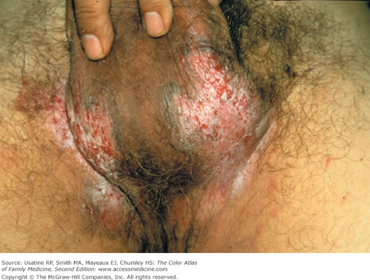
- Perianal Paget disease (Figure 86-2) is associated with underlying colorectal carcinoma in 25% to 35% of cases.
Etiology and Pathophysiology
Risk Factors
- Extramammary Paget disease should be considered in any patient with chronic dermatitis of the groin, vulva, scrotal, or perianal area. Patients with Paget disease of the external genitalia often present with nonresolving eczematous lesions in the groin, genitalia, perineum, or perianal area.7
Diagnosis
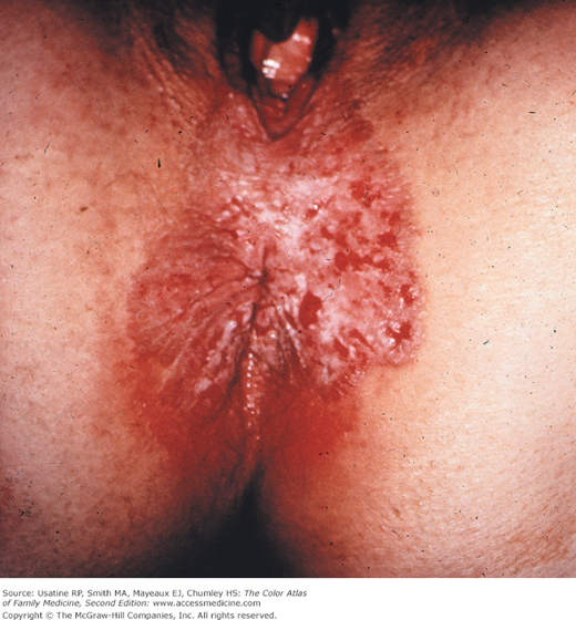
- Lesions present on the vulva as geographic red macules that often appear excoriated or have an eczematoid appearance (Figures 86-1, 86-2, and 86-3). The lesions are often dotted with small, white patches (islands of tissue). Other lesions may be erythematous, eczematous, or leukoplakic plaques (Figure 86-4).2
- Pruritus occurs in approximately 70% of patients.2 Patients may also experience burning, pain, or no symptoms other than the lesion.6
- The lesions are well-demarcated and have slightly raised edges.
- Vulvar Paget disease is similar in gross appearance to Paget disease of the breast.