Patient Story
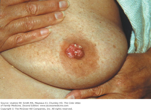
A 62-year-old woman presents with a 6-month history of an eczematous, scaly, rash near her nipple. It is mildly pruritic. On physical examination, the nipple and the areola are involved (Figure 94-1). Also, a hard mass is present in the lateral lower quadrant of the same breast. A 4-mm punch biopsy of the affected area including the nipple demonstrates Paget disease. The mammogram is suspicious for breast cancer at the site of the mass and the patient is referred to a breast surgeon.
Introduction
Epidemiology
- The incidence of Paget disease of the breast is approximately 0.6% in women in the United States, according to National Cancer Institute Surveillance, Epidemiology, and End Results (SEER) data.1 Paget disease, like all breast cancers, is rare in men.
- The peak incidence is between 50 and 60 years of age.2
- It is associated with underlying in situ and/or invasive breast cancer 85% to 88% of the time.3
Etiology and Pathophysiology
- Most patients delay presentation, assuming the abnormality is a benign condition of some sort. The median duration of signs and symptoms prior to diagnosis is 6 to 8 months.2
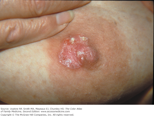
- Presenting symptoms are sometimes limited to persistent pain, burning, and/or pruritus of the nipple (Figures 94-1 and 94-2).
- A palpable breast mass is present in 50% of cases, but is often located more than 2 cm from the nipple–areolar complex.4
- Twenty percent of cases will have a mammographic abnormality without a palpable mass, and 25% of cases will have neither a mass nor abnormal mammogram, but will have an occult ductal carcinoma.
- In less than 5% of cases, Paget disease of the breast is an isolated finding.4
- There are two theories regarding the pathogenesis of Paget disease of the breast, the choice of which affects treatment choices.
- The more widely accepted epidermotropic theory proposes that the Paget cells arise from an underlying mammary adenocarcinoma that migrates through the ductal system of the breast to the skin of the nipple. It is supported by the fact that Paget disease is usually associated with an underlying ductal carcinoma, and both Paget cells and mammary ductal cells usually express similar immunochemical staining patterns and molecular markers. This could mean that there is a common genetic alteration and/or a common progenitor cell for both Paget cells and the underlying ductal carcinoma.
- The less widely accepted transformation theory proposes that epidermal cells in the nipple transform into malignant Paget cells, and that Paget disease of the breast represents an independent epidermal carcinoma in situ. It is supported by the fact that there is no parenchymal cancer identified in a small percentage of cases, and underlying breast carcinomas are often located at some distance to the nipple. Most pathologists disagree with the transformation theory.
- The more widely accepted epidermotropic theory proposes that the Paget cells arise from an underlying mammary adenocarcinoma that migrates through the ductal system of the breast to the skin of the nipple. It is supported by the fact that Paget disease is usually associated with an underlying ductal carcinoma, and both Paget cells and mammary ductal cells usually express similar immunochemical staining patterns and molecular markers. This could mean that there is a common genetic alteration and/or a common progenitor cell for both Paget cells and the underlying ductal carcinoma.
Diagnosis
- Paget disease of the breast presents clinically in the nipple-areolar complex as a dermatitis that may be erythematous, eczematous, scaly, raw, vesicular, or ulcerated (Figures 94-1, 94-2, 94-3, and 94-4). The nipple is usually initially involved, and the lesion then spreads to the areola. Spontaneous improvement or healing of the nipple dermatitis can occur and should not be taken as an indication that Paget disease is not present. The diagnosis is made by finding malignant, intraepithelial adenocarcinoma cells on pathology. Rarely, nipple retraction is found.
- Pain, burning, and/or pruritus may be present or even precede clinically apparent disease develops on the skin.
Stay updated, free articles. Join our Telegram channel

Full access? Get Clinical Tree


