Overview of Digestion and Absorption


Overview of Digestion and Absorption


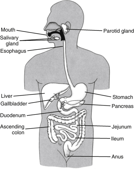
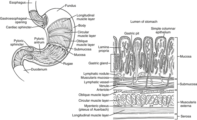
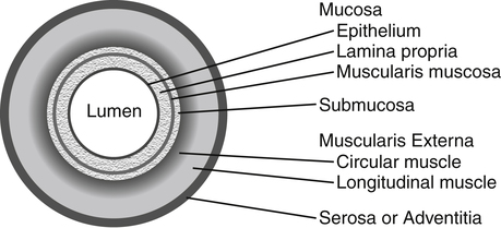
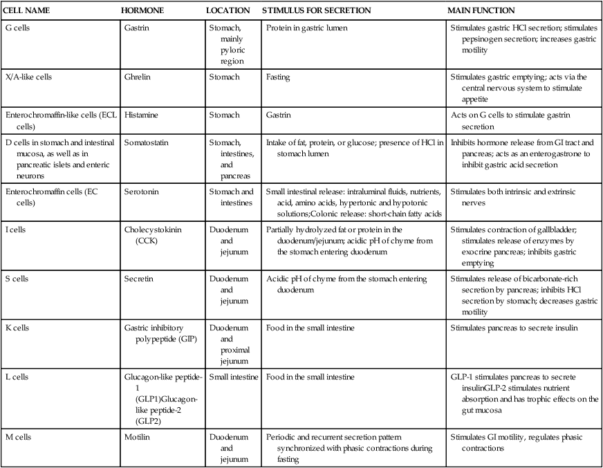
Alan B.R. Thomson, MD, PhD, and Patrick Tso, PhD


Most foodstuffs are ingested in forms that are unavailable to the body and must be broken down into smaller molecules before they can be absorbed into the circulation. The gastrointestinal (GI) tract is the system that carries out the functions of ingestion, digestion, and absorption. The major functions of the GI tract are to digest complex molecules in foods (making them absorbable) and to absorb simple nutrients, including monosaccharides, monoacylglycerols, fatty acids, amino acids, vitamins, minerals, and water. In addition, the GI tract serves excretory, secretory, endocrine, and protective functions.


General Structure and Function of the Gi Tract


The GI tract extends from the mouth to the anus and consists of a long muscular tube with a continuous lumen that opens to the exterior at both ends (Figure 7-1). It has openings for the entry of secretions from the salivary glands, the liver, and the pancreas. The GI system includes the oral cavity, pharynx, esophagus, stomach, small intestine, large intestine, and rectum, as well as accessory organs (salivary glands, pancreas, liver, and gallbladder) that provide essential secretions.


Basic Anatomical Structure of the Gi Tract


Once past the oral cavity, most of the digestive tract has a distinct anatomical structure that is typical of tubular organs (Figure 7-2). Although there are variations along the GI tract, its wall generally comprises four basic tissue layers surrounding the lumen.


Serosa or Adventitia


The fourth basic layer is either the serosa or the adventitia. The intraperitoneal organs of the digestive tract (i.e., upper 5 cm of duodenum, jejunum, ileum, cecum, appendix, transverse colon, sigmoid colon, and upper part of the rectum) are freely suspended in the peritoneal cavity and have a serosa, which is a thin layer of loose connective tissue covered by simple squamous epithelium. The serosa is basically an extension of the peritoneum that lines the wall of the peritoneal cavity and holds the abdominal organs in place. The serosa secretes a watery lubricant that allows parts of the gut to move smoothly over each other. Other parts of the GI tract that are outside of the peritoneal cavity, such as the esophagus and lower part of the rectum, have an outermost connective tissue layer known as adventitia. The retroperitoneal organs (i.e., lower part of the duodenum, ascending and descending colon, and middle part of the rectum) are bound to the posterior abdominal wall by adventitia and covered on their free anterior surfaces by serosa.


General Mechanisms for Regulation of Gi Tract Function


The digestion and absorption of nutrients are both neurally and hormonally regulated.


Intrinsic and Extrinsic Nervous Systems


The GI tract is innervated by both a local intrinsic nervous system and the extrinsic autonomic nervous system. The intrinsic enteric nervous system is responsible for much of the neural regulation of GI motility and function. The enteric nervous system comprises the myenteric plexus, which lies between the muscular layers of the gut and is involved in coordinating the movement of food through the GI tract, and the Meissner plexus, which lies in the submucosa that is under the inner mucosal layer of the gut and is involved in the control of GI secretions and blood flow in the gut wall. The enteric nervous system has both sensory and motor neurons that detect changes in the gut (e.g., as a result of ingesting food or drink) and regulate secretion and motility.


The parasympathetic and sympathetic arms of the autonomic nervous system function along with the enteric nervous system to regulate gut function. In general, parasympathetic stimulation promotes digestion and absorption by increasing GI secretion, increasing muscle tone and peristalsis, and relaxing GI sphincters and blood vessels. On the other hand, sympathetic stimulation has the opposite effects, leading to decreased secretion, decreased muscle tone and movement, and constriction of sphincters and blood vessels. The major parasympathetic supply to the GI tract is via the vagus and pelvic nerves, whose nerve endings in the GI tract release acetylcholine, whereas the major sympathetic nerve endings secrete mainly norepinephrine.


The arrangement of the enteric nervous system and its connections with the sympathetic and parasympathetic systems support several types of GI reflexes that are important in GI control. These reflexes may be integrated entirely within the gut wall enteric nervous system or may involve afferent signals from the receptors in the GI tract that travel back to the central nervous system to trigger efferent output to the same or different parts of the GI tract.


GI Hormones


GI hormones are secreted by enteroendocrine cells located throughout the GI tract (Table 7-1). These enteroendocrine cells do not form endocrine glands but rather secrete GI hormones that exert local effects on the GI tract via paracrine and autocrine actions. They also affect other tissues via endocrine actions after entering the circulation. GI hormones play important roles in regulating GI functions, such as gut motility and the secretion of hydrochloric acid (HCl). The actions of these hormones on other tissues, such as brain, pancreas, liver, and gallbladder, also affect the regulation of the GI tract by these tissues.


TABLE 7-1


The Endocrine Cells of the GI Tract


CELL NAME HORMONE LOCATION STIMULUS FOR SECRETION MAIN FUNCTION
G cells Gastrin Stomach, mainly pyloric region Protein in gastric lumen Stimulates gastric HCl secretion; stimulates pepsinogen secretion; increases gastric motility
X/A-like cells Ghrelin Stomach Fasting Stimulates gastric emptying; acts via the central nervous system to stimulate appetite
Enterochromaffin-like cells (ECL cells) Histamine Stomach Gastrin Acts on G cells to stimulate gastrin secretion
D cells in stomach and intestinal mucosa, as well as in pancreatic islets and enteric neurons Somatostatin Stomach,
intestines, and
pancreas
Intake of fat, protein, or glucose; presence of HCl in stomach lumen Inhibits hormone release from GI tract and pancreas; acts as an enterogastrone to inhibit gastric acid secretion
Enterochromaffin cells (EC cells) Serotonin Stomach and intestines Small intestinal release: intraluminal fluids, nutrients, acid, amino acids, hypertonic and hypotonic solutions;Colonic release: short-chain fatty acids Stimulates both intrinsic and extrinsic nerves
I cells Cholecystokinin (CCK) Duodenum and jejunum Partially hydrolyzed fat or protein in the duodenum/jejunum; acidic pH of chyme from the stomach entering duodenum Stimulates contraction of gallbladder; stimulates release of enzymes by exocrine pancreas; inhibits gastric emptying
S cells Secretin Duodenum and jejunum Acidic pH of chyme from the stomach entering duodenum Stimulates release of bicarbonate-rich secretion by pancreas; inhibits HCl secretion by stomach; decreases gastric motility
K cells Gastric inhibitory polypeptide (GIP) Duodenum and proximal jejunum Food in the small intestine Stimulates pancreas to secrete insulin
L cells Glucagon-like peptide-1 (GLP1)Glucagon-like peptide-2 (GLP2) Small intestine Food in the small intestine GLP-1 stimulates pancreas to secrete insulinGLP-2 stimulates nutrient absorption and has trophic effects on the gut mucosa
M cells Motilin Duodenum and jejunum Periodic and recurrent secretion pattern synchronized with phasic contractions during fasting Stimulates GI motility, regulates phasic contractions


image


GI, Gastrointestinal; HCI, hydrochloric acid.


Basic Functions of the Gi Tract


The major functions of the GI tract are digestion and absorption.


Digestion


Both mechanical (physical) and enzymatic (chemical) processes are involved in digestion. Mechanical digestion includes the chewing of food and the muscular movements of the stomach and intestines that break food particles into smaller pieces, thus exposing a greater surface area of the food to digestive enzymes. Mechanical digestion also mixes food with various secretions from the GI tract and accessory organs, which facilitates the action of digestive enzymes on the food particles. Chemical digestion is defined as the breakdown of food by enzymes. These enzymes either are secreted into the lumen of the GI tract by glandular cells in the mouth, chief cells in the stomach, or exocrine cells of the pancreas; or they are resident enzymes of the brush border (luminal) membrane or the cytoplasm of mucosal cells of the small intestine. Digestion also involves the effect of gastric acid to denature proteins. Digestion occurs before nutrients can cross the absorptive cells to enter the interstitial fluid/circulatory system.


Absorption


Absorption is the movement of nutrients, including water and electrolytes, across the mucosal epithelial lining facing the lumen of the GI tract into the lamina propria (interstitium), where they enter either the blood or the lymph. Nutrient absorption includes both transcellular (through the epithelial absorptive cells) and paracellular (between the epithelial cells) mechanisms. Most nutrient absorption occurs in the small intestine, which has specialized absorptive cells that effect transcellular uptake. Substances pass from the intestinal lumen into the absorptive cells and then out of the absorptive cells to the extracellular compartment. The processes responsible for movement across the luminal membrane of the absorptive cells are often quite different from those responsible for movement across the basolateral or contraluminal cell membranes to the lamina propria. Paracellular uptake by movement through pores between cells may accomplish absorption of water, ions, and some other small molecules. Once nutrients have entered the lamina propria, they enter either the capillaries (into the blood) or the lacteals (into the lymph).


Other Functions of the GI Tract


In the context of digestion, the GI tract has many secretory functions that help to moisten and lubricate the food, adjust the pH of the GI tract contents, and supply some of the enzymes needed for the luminal digestion process. The GI tract also has important endocrine functions because it contains many enteroendocrine cells that release hormones into the bloodstream. Many of these regulate GI tract function, and some regulate food intake. Excretion is another important function of the GI tract. Undigested food material, bacterial mass, and metabolic wastes excreted by the liver in the bile are all excreted by the GI tract in the feces. In addition, the GI tract is an important part of the immune system. Immunoreactive cells in the lamina propria and submucosa of the GI tract, the layer of mucus covering the epithelium, the tight junctions between the epithelial cells of the mucosa, and lysozyme secretion by Paneth cells in the small intestine all serve to protect the body from pathogens that can enter the body through the GI tract.


The Upper Gi System


Sometimes the GI tract is divided into upper and lower parts. The upper GI regions include the oral cavity, the esophagus, and the stomach. The lower GI tract includes the small intestine, the large intestine, and the rectum. Sometimes the first part of the small intestine, the duodenum, is divided into two parts based upon arterial supply or embryology, with the upper region of the duodenum considered part of the upper GI tract.


The Oral Cavity


The process of digestion and absorption begins with the ingestion of food. A number of factors are involved in regulation of food intake, and the complex process of this regulation is discussed in Chapter 22. In the oral cavity, chewing involves the cutting and grinding of food by the teeth and the crushing of the food bolus into smaller particles. The process of chewing also mixes food with saliva. Secretion of saliva by the salivary glands is stimulated by the parasympathetic nervous system in response to the sight, smell, and taste of food and mechanical stimulation of the oral cavity.


Saliva has many functions, including partial digestion of starch by salivary amylase; antibacterial activity (due to the presence of thiocyanate, lactoferrin, and lysozyme); moistening of the mouth to facilitate speech, chewing, and swallowing; neutralization of acids (due mainly to presence of bicarbonate and carbonic anhydrase in the saliva); moistening and lubricating the bolus of food to facilitate swallowing; and maintaining oroesophageal tissue integrity (due to secretion of epidermal growth factor, which may help to repair the esophagus if it has been damaged, for example by gastric acid). Although salivary amylase is mixed with food in the mouth, most of the digestion by this enzyme is accomplished in the stomach rather than in the oral cavity. The salivary amylase mixed in the food bolus continues to be active in the interior of the bolus for a period of time after the food has been swallowed. Once the food bolus is broken up and the amylase is inactivated by the acidic secretions of the stomach, its action ends. In some species, lingual lipase is secreted by glands in the tongue, and this enzyme is most active in the acidic environment of the gastric lumen. Secretion of lingual lipase is insignificant in humans, but the stomach of humans does secrete a lipase that has similar acid lipase activity.


Swallowing


Swallowing is the process by which the food bolus passes from the mouth to the pharynx and on through the esophagus into the stomach. Swallowing begins with the oral phase that is under voluntary neuromuscular control. This initial voluntary action, however, triggers the pharyngeal and esophageal phases, which are under involuntary control by the autonomic nervous system. The swallowing reflex is initiated by touch receptors in the pharynx as the food bolus is pushed to the back of the mouth by the tongue. This swallowing reflex consists of a series of highly coordinated responses that include interruption of breathing, movement of the soft palate to close the entrance to the nasal cavities, movement of the tongue to close the exit from the pharynx back into the mouth, closure of the larynx by the epiglottis so that food does not enter the trachea, contraction of the pharynx to force the bolus into the esophagus, and peristaltic movements in the esophagus that carry the food to the stomach.


The Esophagus


The esophagus has a typical tubular organ structure with a sphincter at each end (i.e., at the junctions of the esophagus with the pharynx and the stomach). The mucosa of the esophagus is distensible to accommodate the swallowing of a fairly large bolus of food and hence has a somewhat puckered appearance when the underlying muscle has not been stretched. The mucosal epithelium is a stratified squamous epithelium that is resistant to abrasion. Submucosal glands secrete mucus. In humans, the inner circular and outer longitudinal muscle layers are primarily composed of skeletal muscle fibers in the upper portion of the esophagus and of smooth muscle in the lower part of the esophagus near the junction with the stomach. The food bolus can move through the approximately 25-cm-long esophagus and enter the stomach in less than 10 seconds.


Reflexes initiated during swallowing relax the upper esophageal sphincter to let food pass, after which various muscles of the pharynx as well as peristalsis by the muscles of the esophagus sequentially push the bolus of food through the esophagus. Coordinated contractions of the esophageal muscles produce the waves of peristalsis that propel the bolus through the esophagus. The lower esophageal sphincter relaxes before the contraction wave, allowing food to enter the stomach. The lower esophageal sphincter then closes to prevent the reflux of stomach acid into the esophagus.


When the lower esophageal sphincter does not close properly, the acidic stomach contents rise up into the esophagus. This is known as acid reflux or acid regurgitation. During pregnancy, the lower esophageal sphincter may be more relaxed than usual, and there may be increased pressure on the stomach as a result of the growing baby. This may allow the reflux of acid into the esophagus, giving the feeling of heartburn and regurgitation. Smoking and obesity are also factors that contribute to frequent acid reflux. Certain foods, such as alcohol, chocolate, and peppermints, may worsen symptoms because they relax the lower esophageal sphincter and slow gastric emptying. Occasional gastroesophageal reflux is common, but persistent reflux that occurs more than twice per week is considered more serious because the stomach acid can damage and inflame the esophageal mucosa. Gastroesophageal reflux disease may occur because of weak contraction or abnormal relaxation of the lower esophageal sphincter, a hiatal hernia in which a small portion of the stomach pushes up through the diaphragm, or weak muscle contractions in the esophagus.


The Stomach


The food bolus exits the lower esophageal sphincter through the cardiac orifice, the opening that connects the cardia region of the stomach to the esophagus. Vagal reflexes initiated by the cephalic phase of eating (i.e., by the sight, smell, thought, or taste of food) inhibit contractile activity in the proximal stomach even when the stomach is empty, and the entry of food into the stomach promotes relaxation of the cardia of the stomach. Thus the stomach muscles relax in preparation for the entrance of food and in response to the entrance of food. When relaxed and empty, the adult human stomach has a volume of about 0.05 L, but it normally expands to hold about 1 L of food and liquid and can hold up to 2 L or more with associated discomfort.


Functions of the Stomach


The stomach serves several functions in digestion and absorption. First, it temporarily stores the swallowed food and liquid until it is passed to the intestines. Second, the stomach secretes HCl, enzymes or zymogens needed for initiating digestion, endocrine hormones that regulate the process of food assimilation, and intrinsic factor that is essential for vitamin B12 absorption in the small intestine. Third, the stomach mixes up the food, liquid, and digestive juice produced by the stomach and then macerates this mixture into a semiliquid state. This semiliquid mass of partly digested food is called chyme. Fourth, the stomach regulates the rate of entry of chyme into the small intestine. Although food undergoes substantial physical and chemical modifications in the stomach, little absorption occurs. Some water, salts, and lipid-soluble substances such as ethanol and short-chain fatty acids are absorbed, however.


Basic Anatomy of the Stomach


The stomach has four gross anatomical regions: cardia, fundus, body (or corpus), and pyloric antrum (Figure 7-3). The body, which is the largest part of the stomach, extends from the cardia to the pylorus, the sections that connect with the esophagus and the small intestine, respectively. The fundus is the upper end of the stomach that lies above the cardia and superior to the opening of the esophagus. The pylorus is sometimes described as having two sections: a pyloric antrum that narrows into the pyloric canal. The pyloric canal is surrounded by thickened, circular smooth muscle fibers to form a pyloric sphincter at the opening of the pyloric canal into the duodenum; this sphincter helps control the evacuation of food from the stomach. The lining of the stomach has folds or plaits called rugae, which are most pronounced toward the pyloric end of the stomach. They flatten and disappear as the stomach is filled and distends.


The tubular structure of the stomach wall follows the basic four-layer structure: mucosa, submucosa, muscle layers, and serosa. The mucosa comprises a simple columnar epithelium, which lines the luminal surface of the stomach and the gastric pits; the lamina propria, which contains various glands, depending on the region of the stomach; and a thin smooth muscle layer. The submucosa of the stomach contains no glands. The muscle layers of the stomach include the circular (middle) and longitudinal (outer) layers of muscle found in other parts of the GI system as well as an inner oblique layer that is responsible for creating the motion that churns and physically grinds the food. The serosa consists of connective tissue that is continuous with the peritoneum. The simple columnar epithelial cells that form the luminal surface layer of the stomach are usually referred to as surface mucous cells because they secrete mucus. The layer of mucus on the surface of the mucosal epithelium is important in protecting the stomach lining from damage by acid and proteolytic enzymes secreted by the gastric glands. In fact, the pH of the mucus layer remains nearly neutral because of secretion of bicarbonate by the underlying mucosa, whereas the pH becomes very acidic (pH ~1 to 3) in the stomach lumen due to HCl secretion from the gastric pits when the gastric glands are stimulated.


Gastric Pits and Gastric Glands


The gastric mucosa contains many minute depressions called gastric pits that extend into the mucosal layer. Glands that contain a mixed population of cell types are associated with these gastric pits. The cell types and secretions of the gastric glands vary somewhat by the region of the stomach in which they are located. The glands extend deep into the mucosa layer, surrounded by the connective tissue of the lamina propria, and they open into the base of the gastric pits. Although connective tissue separates the individual glands, most of the volume of the gastric mucosa is occupied by secretory cells, primarily the parietal and chief cells.


Gastric glands contain a mixture of several cell types, including mucoid cells, parietal cells, chief cells, and enteroendocrine cells. The mixed secretions of the exocrine gastric glands are referred to as the gastric juice. The main components of human gastric juice are mucus, secreted by mucoid cells; HCl and intrinsic factor, secreted by parietal cells; and pepsinogens and gastric lipase, secreted by chief cells, along with water, electrolytes, and bicarbonate. Gastric juice is secreted in response to the vagal stimulation associated with eating (e.g., in response to taste and smell) and in response to gastrin released by enteroendocrine cells in the stomach.


The products of the enteroendocrine cells are not secreted into the lumen of the gastric pit and are not a part of the gastric juice. The enteroendocrine secretions may act locally via paracrine mechanisms or be taken up from the extracellular fluid into the bloodstream to circulate to various tissues as endocrine hormones. Vagal reflexes initiated during the initial phase of eating, as well as the lower luminal acidity and distention caused by the entrance of food into the stomach, stimulate release of hormones, including gastrin, by the enteroendocrine cells of the stomach. Protein in food is also a potent stimulator of gastrin release. Gastrin (from G cells) stimulates HCl production by parietal cells in the gastric glands. The main mechanism of gastrin’s action is thought to involve enterochromaffin-like (ECL) cells. ECL cells are stimulated by gastrin to release histamine, and it is thought that the stimulation of parietal cells is mainly due to histamine binding secondary to gastrin’s effects on histamine release. Gastrin secretion is inhibited by feedback from acid within the lumen of the stomach. This feedback mechanism regulates the amount of gastrin released and therefore the amount of acid secreted in response to a meal.


Ghrelin is another hormone released by the stomach. The release of ghrelin is stimulated by fasting and is suppressed by the ingestion of food. Ghrelin stimulates gastric emptying and acts via the central nervous system to stimulate appetite.


Hydrochloric Acid Secretion and Function


The parietal cells are the source of gastric HCl. Parietal cells contain secretory channels called canaliculi, from which the gastric acid is secreted into the lumen of the stomach. Gastric acid secretion happens in several steps. First, chloride and hydrogen ions are secreted separately from the cytoplasm of parietal cells and mixed in the canaliculi. H+ ions are generated within the parietal cell from dissociation of H2O. The OH ions formed in this process combine with CO2, in a reaction catalyzed by carbonic anhydrase, to form HCO3. The HCO3 is transported out of the cell in exchange for Cl ions. Thus the parietal cell now has a supply of both H+ and Cl ions for secretion. Cl and K+ ions are transported into the lumen of the canaliculus by conductance channels. H+ ions are then pumped out of the cell in exchange for K+ by the membrane H+,K+-ATPase (proton pump). The accumulation of osmotically active ions in the canaliculus causes water to diffuse outward from the parietal cell, generating a secretion rich in HCl. The bicarbonate that was transported out of the parietal cells causes the venous blood leaving the stomach to temporarily be more alkaline than the arterial blood delivered to it. This phenomenon is known as the alkaline tide. Gastric acid is then secreted into the lumen of the gastric gland and gradually reaches the main stomach lumen.


The resulting acidic environment (pH ~1 to 3) in the stomach lumen favors the denaturation (unfolding) of proteins, which makes the peptide bonds in the proteins more accessible to proteolytic enzymes. The acidic environment is also necessary to activate the pepsinogens secreted by the chief cells (i.e., to convert pepsinogens to pepsins via autocatalytic cleavage) and to promote the enzymatic activity of the pepsins, which exhibit optimal activity and stability at an acidic pH of approximately 2. Additionally, the gastric acidity destroys many microorganisms that enter the GI tract via the oral cavity. Some organisms such as Helicobacter pylori can survive the acidic pH of the lumen long enough to burrow through the mucoid lining of the stomach to find a niche close to the epithelial cell layer. H. pylori infection is a common and easily treatable cause of peptic ulcers.

Stay updated, free articles. Join our Telegram channel

Feb 26, 2017 | Posted by in PHARMACY | Comments Off on Overview of Digestion and Absorption

Full access? Get Clinical Tree

Get Clinical Tree app for offline access