Composed of enlarged cells with abundant clear to vacuolated-appearing cytoplasm
• Chondroid melanoma
Malignant epithelioid cells associated with prominent myxochondroid-appearing matrix
• Osteoid melanoma
Malignant epithelioid cells associated with prominent osteoid-appearing material
• Myxoid melanoma
Atypical epithelioid cells scattered in prominent myxoid matrix
• Small cell melanoma
Nests and sheets of small, hyperchromatic-staining cells with scant cytoplasm (high nuclear:cytoplasmic ratio)
• Rhabdoid melanoma
Proliferation of markedly enlarged and atypical-appearing melanocytes with abundant eosinophilic-staining cytoplasm
Top Differential Diagnoses
• Nevi
• Chondroma or other tumors with cartilaginous differentiation
• Osteoma or other tumors with osseous differentiation
• Benign cutaneous myxoid proliferations
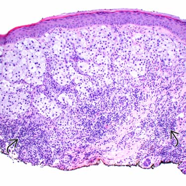
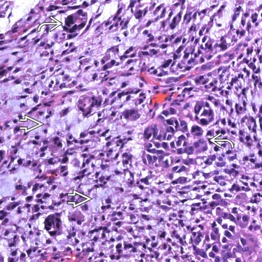
Balloon Cell Melanoma Balloon cell melanoma is composed of nests of large, pale- to clear-staining cells with abundant vacuolated cytoplasm (mimicking sebocytes). Note the associated lymphocytic infiltrate .
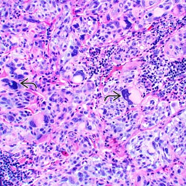
Myxoid Melanoma High magnification of myxoid melanoma shows many single tumor cells and small nests floating in a prominent mucinous stroma .
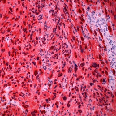
Spindle Cell/Pleomorphic Melanoma This is a case of spindle cell/pleomorphic melanoma, which can mimic spindle cell carcinoma, atypical fibroxanthoma, and pleomorphic sarcoma (as well as other sarcomas). Note the frankly pleomorphic, bizarre-appearing tumor giant cells .
S100 Immunohistochemical Stain in Spindle Cell/Pleomorphic Melanoma S100 (red chromogen) shows strong and diffuse staining in this case of spindle cell/pleomorphic melanoma, essentially excluding the other tumors in the differential diagnosis.
TERMINOLOGY
Definitions
• Rare variants of melanoma, including clear cell/balloon cell, chondroid, osteoid, myxoid, and rhabdoid melanoma
ETIOLOGY/PATHOGENESIS
Environmental Exposure
• Related to UV exposure in most cases
CLINICAL ISSUES
Epidemiology
• Incidence
Very rare tumors
• Age
Typically elderly patients
Site
• Sun-damaged skin, typically head and neck region, upper trunk, forearms
Presentation
• Usually present as papule or nodule with irregular, asymmetric borders
Treatment
• Surgical approaches
Complete and wide excision, with clinical margins based on Breslow depth
Prognosis
• Depends on typical melanoma prognostic features, such as Breslow depth, ulceration, mitotic count, and presence of perineural or angiolymphatic invasion
• Rhabdoid melanoma reportedly shows aggressive course in most cases
MICROSCOPIC
Histologic Features
• Depends on histologic variant
• Most cases often show areas of conventional-appearing melanoma &/or overlying melanoma in situ
• Clear cell/balloon cell melanoma
Enlarged cells with abundant clear- to vacuolated-appearing cytoplasm
– May be due to glycogen
May appear deceptively bland in some cases
• Chondroid melanoma
Malignant epithelioid cells associated with myxochondroid-appearing matrix
Often shows areas of overlying melanoma in situ or more conventional melanoma with nesting
Only gold members can continue reading. Log In or Register to continue


 .
.
 floating in a prominent mucinous stroma
  .
.
 .
.