Introduction
There are a number of bullous diseases other than pemphigus and bullous pemphigoid that are important to recognize. Porphyria cutanea tarda is a porphyria that has no extracutaneous manifestations (Figures 186-1, 186-2, and 186-3). Dystrophic epidermolysis bullosa belongs to a family of inherited diseases where blister formation can be caused by even minor skin trauma. PLEVA (pityriasis lichenoides et varioliformis acuta) is a minor cutaneous lymphoid dyscrasia that can appear suddenly and persist for weeks to months. Dermatitis herpetiformis is a recurrent eruption that is usually associated with gluten and diet-related enteropathies.
Porphyria Cutanea Tarda
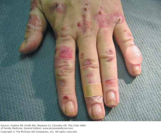
A middle-aged woman presented with tense blisters on the dorsum of her hand (Figure 186-1). One bulla was intact and the others had ruptured, showing erosions. Work-up showed elevated porphyrins in the urine (which fluoresced orange-red under a Wood lamp) and the patient was diagnosed with porphyria cutanea tarda.
- Porphyria cutanea tarda (PCT) occurs mostly in middle-aged adults (typically 30 to 50 years of age) and is rare in children.
- It is especially likely to occur in women on oral contraceptives and in men on estrogen therapy for prostate cancer.1
- Alcohol, pesticides, and chloroquine have been implicated as chemicals that induce PCT.1
- PCT is equally common in both genders.
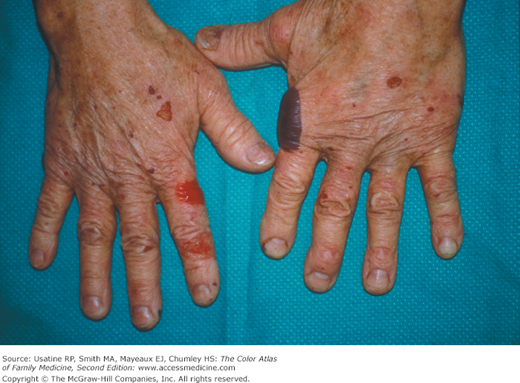
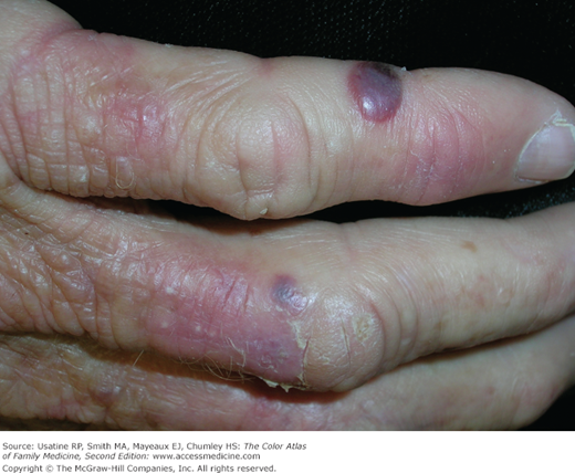
- There is an increased incidence of PCT in persons with hepatitis C (Figures 186-2 and 186-3).
- The porphyrias are a family of illnesses caused by various metabolic derangements in the metabolism of porphyrin, the chemical backbone of hemoglobin. Whereas the other porphyrias (acute intermittent porphyria and variegate porphyria) are associated with well-known systemic manifestations (abdominal pain, peripheral neuropathy, and pulmonary complications), PCT has no extracutaneous manifestations. Photosensitivity is seen (as with variegate porphyria). PCT is associated with a reduction in hepatic uroporphyrin decarboxylase.
The classic presentation is that of blistering (vesicles and tense bullae) on photosensitive “fragile skin” (similar to epidermolysis bullosa). Scleroderma-like heliotrope suffusion of the eyelids and face may be seen. As the blisters heal, the skin takes on an atrophic appearance. Hypertrichosis (especially on the cheeks and temples) is also common and may be the presenting feature.
Classically, the dorsa of the hands are affected (Figures 186-1, 186-2, and 186-3). Facial suffusion (heliotrope) may be seen along with hypertrichosis of the cheeks and temples.
The diagnosis can be confirmed by the orange-red fluorescence of the urine when examined under a Wood lamp. Increased plasma iron may be seen (associated with increased hepatic iron in the Kupffer cells). Diabetes is said to occur in 25% of individuals.
- Twenty-four-hour urine collection for porphyrins—These will be elevated in PCT.
- Skin biopsy may help confirm PCT if the other information is not clear.
- Once the diagnosis is made, secondary causes of PCT should be investigated:
- Serum for ferritin, iron, and iron-binding capacity to look for hemochromatosis.
- Order liver function tests and if abnormal order tests for hepatitis B and C.
- Consider α-fetoprotein and liver ultrasound if considering cirrhosis and/or hepatocellular carcinoma.
- Order an HIV test if risk factors are present.
- Serum for ferritin, iron, and iron-binding capacity to look for hemochromatosis.
- The acral vesiculobullous lesions may suggest nummular or dyshidrotic eczema. In younger individuals, the acral blistering may suggest epidermolysis bullosa. The lesions may also suggest erythema multiforme bullosum. The heliotrope suffusion may suggest dermatomyositis and the atrophic changes may suggest systemic sclerosis.
- If the onset is associated with alcohol ingestion, estrogen therapy, or exposure to pesticides, reducing exposure is warranted.2
- Phlebotomy of 500 mL of blood weekly until the hemoglobin is decreased to 10 g is associated with biochemical and clinical remission within a year.1
- Low-dose chloroquine can help maintain remissions, whereas high-dose chloroquine can exacerbate the illness.1
- Periodic clinical follow-up until remission is achieved is necessary along with constant education and reinforcement of the need to avoid precipitants.
Epidermolysis Bullosa
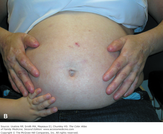
A 34-year-old pregnant woman presents with active blistering in her axilla and past history revealed that she lost her fingernails and toenails (Figure 186-4A) as a young child. She was diagnosed as a child with recessive dystrophic epidermolysis bullosa. None of her children had been affected because her husband was neither affected nor a carrier (Figure 186-4B). A topical steroid ointment helped relieve the pain and calm the blistering in her axilla.
Figure 186-4
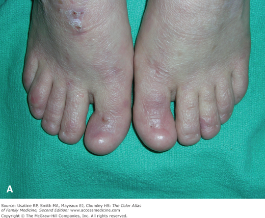
A. Recessive dystrophic epidermolysis bullosa with loss of all her toenails as a young child. B. The same woman showing complete loss of her fingernails and the pregnant abdomen. Her daughter is touching the belly and does not have the disease and, therefore, has normal fingers. (Courtesy of Richard P. Usatine, MD.)
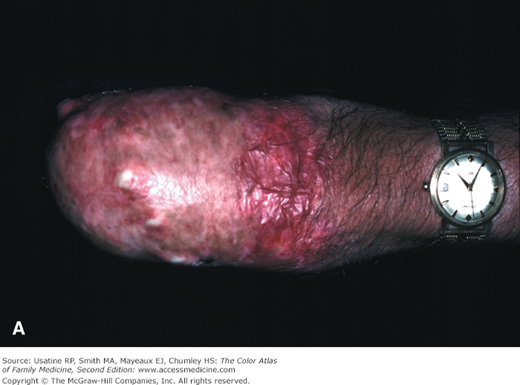
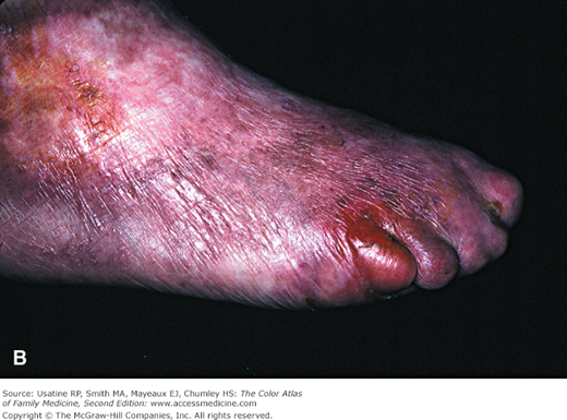
- Dystrophic epidermolysis bullosa belongs to a family of inherited diseases characterized by skin fragility and blister formation caused by minor skin trauma.3 There are autosomal recessive and autosomal dominant types, the severity of this disease may vary widely. Onset is in childhood and in later years severe dystrophic deformities of hands and feet are characteristic (Figure 186-5). Malignant degeneration is common, especially squamous cell carcinoma, in sun-exposed areas.
Figure 186-5
Severe recessive dystrophic epidermolysis bullosa in a 53-year-old Asian man. A. Complete loss of fingers from the disease on his hands. This is referred to as the mitten deformity. He has also had multiple squamous cell carcinomas excised from his hands. B. Similar foot deformities with loss of normal toes. (Courtesy of Richard P. Usatine, MD.)
- Dystrophic epidermolysis bullosa has vesiculobullous skin separation occurring at the sub-basal lamina level, as opposed to junctional epidermolysis bullosa, which blisters at the intralamina lucida layer, and epidermolysis bullosa simplex (Figure 186-6), which blisters at the intraepidermal layer.4,5
Stay updated, free articles. Join our Telegram channel

Full access? Get Clinical Tree


