Osteoma Cutis
David S. Cassarino, MD, PhD
Key Facts
Etiology/Pathogenesis
Primary: May be part of Albright hereditary osteodystrophy or other genetic syndromes
Secondary: Associated with preexisting lesions, such as nevi, tumors, scars, and ruptured cysts
Clinical Issues
Primary cases often present at birth or in early childhood; secondary cases in adults
Microscopic Pathology
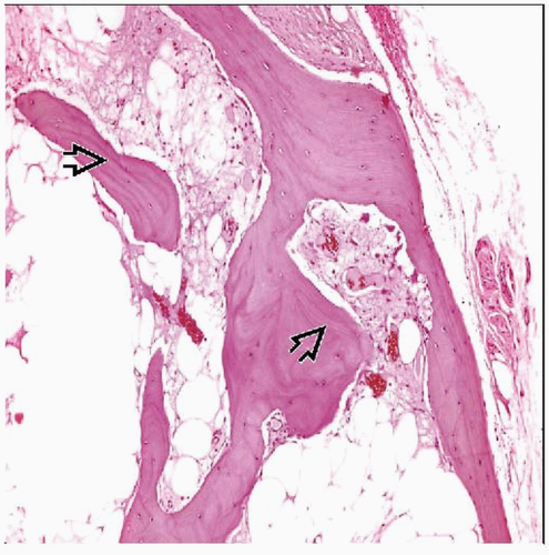
Appearance varies from small spicules to large masses of bone
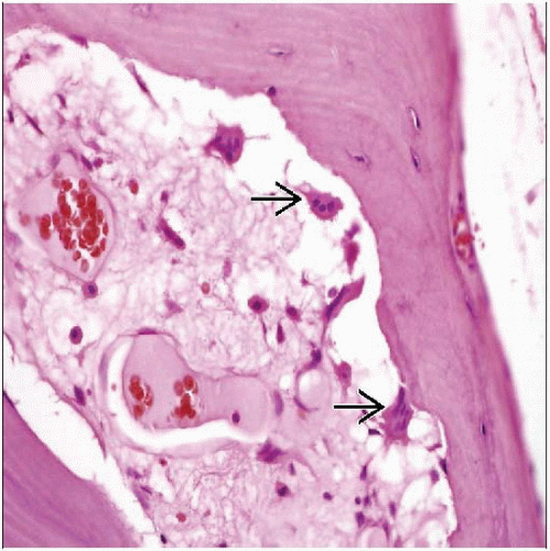
Mature-appearing bone, often with Haversian systems and cement lines
Osteoblastic activity may be present (especially in Albright syndrome), osteoclasts are less common
Typically located in deep dermis &/or subcutaneous tissue
TERMINOLOGY
Synonyms
Primary cutaneous osteoma cutis
Metaplastic (secondary) ossification
Cutaneous ossification
Definitions
Primary or secondary cutaneous bone formation
ETIOLOGY/PATHOGENESIS
Primary
Often genetic or developmental in origin, presents at early age
May be part of Albright hereditary osteodystrophy or other genetic syndromes
Albright: X-linked dominant condition associated with characteristic facies, mental retardation, basal ganglia calcification, and cataracts
Other rare syndromes include congenital plaquelike osteomatosis, multiple miliary osteomas of face, progressive osseous heteroplasia, and fibrodysplasia ossificans progressiva
Secondary
Associated with preexisting lesions, such as nevi, tumors (including adnexal tumors and basal cell carcinoma), scars, acne, and ruptured cysts
Stay updated, free articles. Join our Telegram channel

Full access? Get Clinical Tree


