208 Old choroiditis
Salient features
Examination
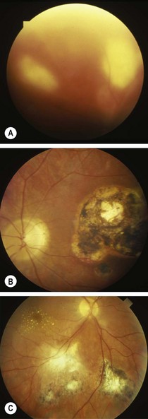
• Old or inactive retinochoroiditis appears as white, well-defined areas of chorioretinal atrophy with pigmented edges (caused by proliferation of retinal pigment epithelium) (Fig. 208.1B)
• The retinal blood vessels pass over the lesions undisturbed.
Stay updated, free articles. Join our Telegram channel

Full access? Get Clinical Tree


