Patient Story
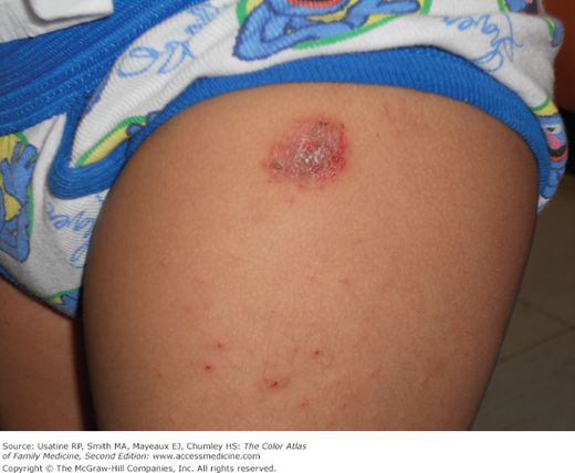
A 2-year-old Hispanic male presents to the clinic with erythematous, round, moist, crusted lesions on the left thigh (Figure 148-1) and right arm. His mother noted several small bumps initially, which developed into coin-shaped lesions over the next few weeks. The child is scratching them but is otherwise healthy. A KOH preparation of the scraping from the lesions did not show fungal structures. The child responds well to treatment with topical midpotency corticosteroids, emollients, and dressing in long-sleeve clothes to prevent scratching the lesions. His nummular eczema resolved in 6 weeks.
Introduction
Nummular eczema (NE) is a type of eczema characterized by circular or oval-shaped scaling plaques with well-defined borders. The term nummular refers to the shape of a coin (Latin for coin is nummus). The lesions are typically multiple and most commonly found on the dorsa of the hands, arms, and legs. It often overlaps with other clinical types of eczema: atopic dermatitis, stasis dermatitis, and asteatotic eczema.1,2
Epidemiology
Etiology and Pathophysiology
Many factors have been reported in association with NE but their role in the etiology and pathogenesis is not well established:
- NE has been viewed as microbial in origin, either secondary to bacterial colonization or hematogenous spread of bacterial toxins,1,3 but an infectious source is not identified in most cases of NE.
- NE is reported to be associated with xerosis of the skin that subsequently weakens the skin barrier function and sensitizes it to environmental allergens.4
- NE is frequently reported in association with contact sensitization to various agents, including nickel, chromate, balsam of Peru, and fragrances. Allergic or chronic contact dermatitis has been frequently reported to manifest as NE on the dorsa of the hands.1,5
- Onset of NE has been reported in association with various medications, including interferon and ribavirin therapy for hepatitis C6,7 and isotretinoin.8 Most of these reports are based on single or limited number of cases.
- Mercury in the dental amalgam was reported to induce NE in two cases with relapsing NE.9
Diagnosis
- Onset is reported to be within days to week. Simultaneous or subsequent development of multiple lesions is often reported.
- Intense pruritus or burning is common.
- Lesions may last months to years without treatment and may be recurrent.
- History of medications, atopy, and exposure to allergens may be helpful to tailor the management of NE.
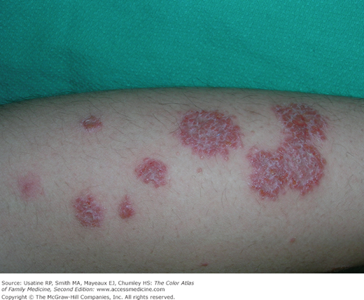
- Primary morphology includes small papules and vesicles that coalesce to form circular to oval shape patches and plaques (Figures 148-2 and 148-3).
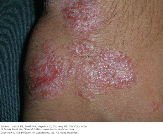
- Secondary morphology includes abrasion and excoriations from scratching (Figure 148-1), weeping and crusting after the vesicles leak (Figures 148-2, 148-3, and 148-4), and scaling and lichenification in more chronic lesions (Figures 148-5 and 148-6). Excessive weeping and crusting may indicate secondary bacterial infection.