Patient Story
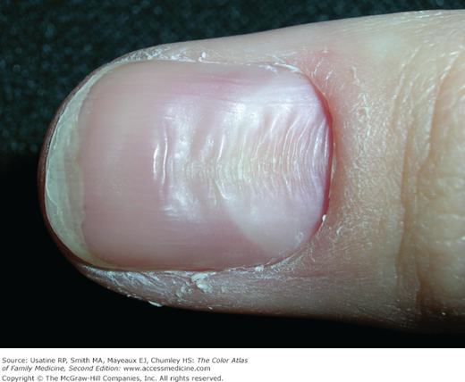
A 28-year-old man is in the office for a work physical and asks about the white streaks on his fingernail (Figure 190-1). He has had them on and off all of his adult life, but recently developed more of them and was concerned he may have a vitamin deficiency. He was reassured that this is a normal nail finding often associated with minor trauma.
Introduction
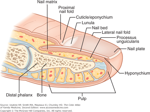
The anatomy of the nail unit is shown in Figure 190-2. The nail unit includes the nail matrix, nail plate, nail bed, cuticle, proximal and lateral folds, and fibrocollagenous supportive tissues. The proximal matrix produces the superficial aspects of the plate, and the distal matrix the deeper portions. The nail plate is composed of hard and soft keratins, is formed via onychokeratinization, which is similar to hair sheath keratinization.1 Most normal nail variants occur as a result of accentuation or disruption of normal nail formation.
Synonyms
- Leukonychia.
- Transverse striate leukonychia.
- Leukonychia punctata.
- White nails.
- Transverse striate leukonychia.
- Longitudinal melanonychia (LM).
- Racial melanonychia in African Americans.
- Nail hypertrophy and onychogryphosis (also known as onychogryposis).
- Ram’s horn nail.
- Oyster-like deformity.
- Lateral nail hypertrophy.
- Thickened toenail.
- Ram’s horn nail.
Epidemiology
Melanonychia often involves several nails and is a more common occurrence in those patients with darker skin types. Among African Americans, benign melanonychia affects up to 77% of young adults and nearly 100% of those age 50 years or older. In the Japanese, LM affects 10% to 20% of adults.1 Nail matrix nevi have been reported to represent approximately 12% of LM in adults and 48% in children.2 The incidences of most other benign nail findings are not well established.
Etiology and Pathophysiology
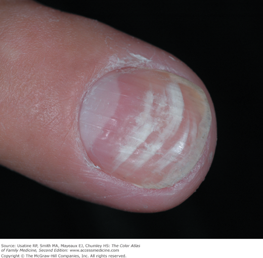
- Leukonychia represents benign, single or multiple, white spots or lines in the nails. Patchy patterns of partial, transverse white streaks (transverse striate leukonychia, see Figure 190-1) or spots (leukonychia punctata, Figure 190-3) are the most common patterns of leukonychia.3 Leukonychia is common in children and becomes less frequent with age. Parents may fear that it represents a dietary deficiency, in particular a lack of calcium, but this concern is almost always unfounded.
- Most commonly, no specific cause for leukonychia can be found. It is usually the result of minor trauma to the nail cuticle or matrix and is the most commonly found nail condition in children.4 When the lesions are caused by overly aggressive manicuring or nervous habit, behavior modification often is helpful. Leukonychia can also be an indirect manifestation of autoimmunity, including alopecia areata or thyroid disease. Histologically, the nail plate contains a greater number of nucleated cells that are associated with lack of cohesion between the corneocytes, producing reflective properties of the nail.
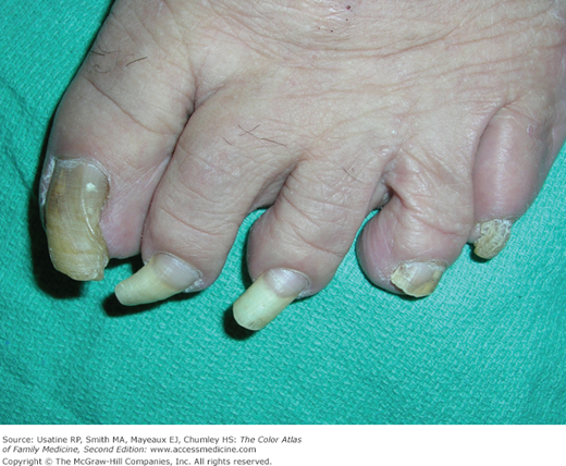
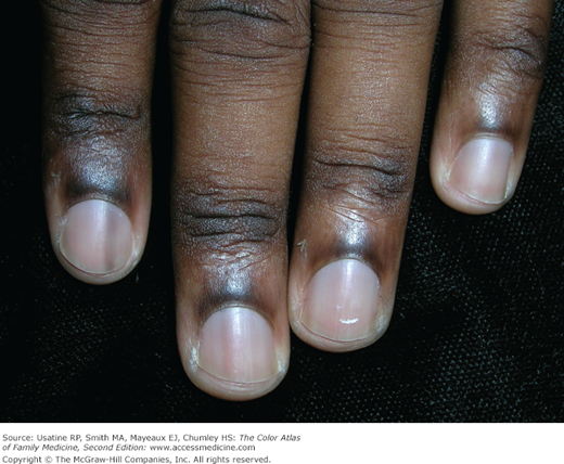
- Longitudinal melanonychia (LM) (Figure 190-4) represents a longitudinal pigmented band in the nail plate. Melanonychia is ultimately caused by melanocyte activation. Causes of nongenetic nail matrix melanocyte activation include drugs, inflammatory processes, trauma, mycosis, systemic diseases, and neoplasms (melanomas).1 LM is often caused by lentigines, benign melanocytic hyperplasia, or nevus of the nail matrix. However, it must be differentiated from subungual melanoma (see Chapter 191, Pigmented Nail Disorders). Benign causes of LM produce melanocytic activation with bands that usually measure 3 to 5 mm or less in width, whereas melanoma tends to produce wider bands. Most lentigines and nevi display a band with a tan-to-brown hue. A benign nail band is generally relatively homogeneous with respect to color and color intensity and if it expands, tends to expand slowly.1
- Nail hypertrophy and onychogryphosis (ram’s horn nail—lateral nail hypertrophy, Figure 190-5) is the development of opaque thickened nails with exaggerated upward, or lateral growth. It may be associated with age, fungal infections and trauma. It can cause pain with pressure.
- Habit-tic deformity (Figures 190-6 and 190-7) is caused by habitual picking of the proximal nail fold. The resulting inflammation induces the nail plate to be wavy and ridged, while its substance remains intact and hard.
- Beau lines are transverse linear depressions in the nail plate (Figures 190-8 and 190-9). They are thought to result from suppressed nail growth secondary to local trauma or severe illness.5 They most commonly appear symmetrically in several or all nails and may have associated white lines. They usually grow out over several months. One may estimate the time since onset of systemic illness by measuring the distance from the Beau line to the proximal nail fold and applying the conversion factor of 6 to 10 days per millimeter of growth.4
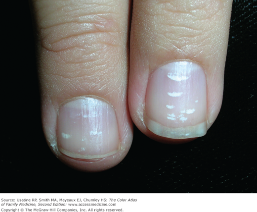
Figure 190-4
Longitudinal melanonychia in multiple fingers in a young adult. These bands of translucent nail pigmentation in multiple fingers are typical of racial longitudinal melanonychia and not suspicious for melanoma. Note the dark pigment on the proximal nail folds represents a pseudo-Hutchinson sign. (Courtesy of Richard P. Usatine, MD.)