Small, Nevoid Appearance Nevoid melanoma is deceptively symmetrical, mimicking an intradermal nevus on low-power examination. The only clue is hypercellularity (blue appearance) and lack of maturation, which should invoke a closer scrutiny.
Numerous Mitotic Figures On high-power examination, numerous mitotic figures are easily found in this nevoid melanoma. This finding is usually surprising given the small size and symmetry on low-power examination.
Deceptive Symmetry Low-power examination shows a deceptively symmetrical-appearing compound, predominantly intradermal proliferation, mimicking a nevus. There is lack of maturation with increasing dermal depth and central hypercellularity.
Lack of Maturation Higher magnification shows that the cells are hyperchromatic staining and do not show definite evidence of maturation with dermal descent. Note also the relatively dense surrounding lymphoid infiltrate . There is an atypical component focally seen.
TERMINOLOGY
Synonyms
• Some cases have been described as “minimal deviation melanoma,” but this term is very controversial and should be avoided
Definitions
• Uncommon variant of melanoma that shows histological features mimicking a nevus
ETIOLOGY/PATHOGENESIS
Environmental Exposure
• Likely solar/UV radiation related, similar to most types of melanoma
CLINICAL ISSUES
Presentation
• Occurs in wide age range
• Can be ubiquitous, but occurs more frequently on back and extremities
• Dome-shaped papule or nodule or verrucous lesion
• Often not recognized clinically as melanoma
Can resemble nevus or basal cell carcinoma
Treatment
• Surgical approaches
Complete excision with clear margins
Sentinel lymph node biopsy often performed for staging, especially if lesion is thicker than 1.0 mm or
– Level IV
– Ulcerated
– Exhibits dermal mitotic figures
Prognosis
• Similar to conventional melanoma
• Determined by AJCC staging, especially depth of invasion (Breslow depth), ulceration, and mitotic activity
MICROSCOPIC
Histologic Features
• At scanning magnification, there are 2 variants
Verrucous or papillated
– Mimics polypoid intradermal melanocytic nevus
Flat or dome-shaped
– Mimics ordinary intradermal or compound melanocytic nevus
• Imparts nevoid appearance on initial examination
Cells mimic type A nevus cells
Can also mimic lymphocytes
• Tightly packed, it forms sheets of relatively banal-appearing nevoid cells
Hypercellular
Only gold members can continue reading. Log In or Register to continue
 and lack of maturation, which should invoke a closer scrutiny.
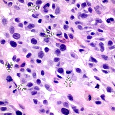
 are easily found in this nevoid melanoma. This finding is usually surprising given the small size and symmetry on low-power examination.
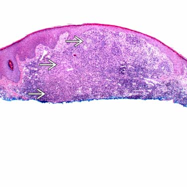
 and central hypercellularity.
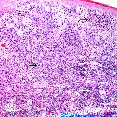
 and do not show definite evidence of maturation with dermal descent. Note also the relatively dense surrounding lymphoid infiltrate
  . There is an atypical component focally seen.


