At this low power, a junctional melanocytic proliferation is evident on the breast of a 25-year-old woman. Most nests  appear at the tips of rete ridges. However, there are apparently displaced nests on the left side of the lesion
  .
.
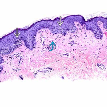
These nests
 appear to be overlying papillary dermal fibroplasia
  and are not laterally displaced or fused as seen in atypical/dysplastic nevi.
Higher power examination exhibits no macronucleoli, vesicular nuclei, or hyperchromasia. Note how the nests
 blend in with epidermal keratinocytes.
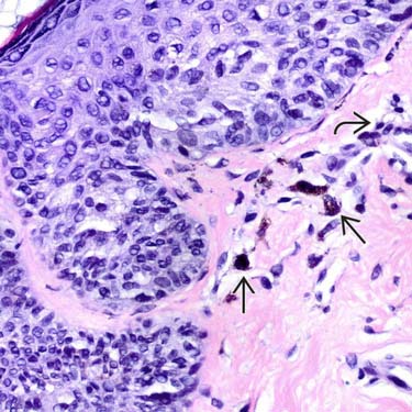
Another focus of the same lesion shows nested melanocytes devoid of cytological atypia. Note the melanin incontinence
 and sparse lymphohistiocytic inflammation
  in the superficial dermis.


MICROSCOPIC
Histologic Features
• Asymmetry and poor peripheral circumscription
 Some nests are randomly scattered along dermoepidermal junction, resembling atypical/dysplastic nevi
Some nests are randomly scattered along dermoepidermal junction, resembling atypical/dysplastic nevi

• Focal upward migration of isolated melanocytes in central part of lesion, especially if traumatized
• Dermal melanocytic component is cytologically bland


Stay updated, free articles. Join our Telegram channel
Full access? Get Clinical Tree
