History
Key features of the history in a patient with neurological disease are shown in Table 6.1.
Table 6.1 Features of the History in Neurological Disease
| Feature | Details | Rationale |
| Basic details | Age, sex, handedness, current and previous occupations | CNS orientation; identify risk of occupational disease |
| Symptoms | Headaches; facial pain; disordered consciousness (‘fits, faints and funny turns’); vertigo; sensory symptoms including numbness, paraesthesiae and pain; motor symptoms including weakness, incoordination, involuntary movements and gait disorders; acute confusional states; memory disorders and symptoms of dementia; speech and language problems; visual disturbances; perceptual problems; sphincter disturbance | Establish pattern of symptoms and their likely causes including relapsing and remitting pattern or progressive problems; establish details of symptoms particularly related to their location and nature, precipitating and relieving factors |
| Past medical history | Cardiovascular disease; diabetes; trauma; inflammatory/immunological disease; infections including HIV/AIDS | Potential causes of neurological disorders |
| Social and family history | Alcohol; smoking; recreational drug use; family history of similar neurological disorders | Potential causes of neurological disease or vascular impairment; potential genetic syndromes |
| Medication | Review all medication | Neurological side effects for many medications |
| Mental state | Assess mental state | Depression is a common feature of chronic neurological syndromes including chronic pain, movement disorders and early dementia |
Examination of the Nervous System
It is important to develop a technique for examination of the nervous system which is rapid and accurate. Analysis of clinical signs is facilitated by knowledge of underlying neuroanatomy shown in the simplified diagrams in this chapter. Examination of the nervous system also requires clear communication with the patient to promote understanding of the sometimes unfamiliar instructions to be given.
Mental State and Higher Cerebral Function
General observation: note
- general appearance and behaviour
- speech
- mood
- abnormal beliefs and/or perceptions
Cognitive Function
Loss of memory for recent events more than for distant events is a feature of organic cerebral disease and an early feature of dementia.
- Use the Mini-Mental State Examination (MME).
- MME consists of 12 questions. Maximum score is 30 and a normal score is 26–30. A score of less than 24 indicates cognitive impairment: 21–25 suggests dementia (likelihood ratio = 5), and 20 or less is highly suggestive of cognitive impairment (likelihood ratio = 8).
Other Tests of Cognitive Function
Concentration: Serial Sevens
Ask the patient:
- to subtract 7 from 100, then seven from the answer and so on
- to remember a series of numbers forward and backward: most people can remember five or more forward and four or more backward
- to repeat a complex sentence, e.g: ‘The one thing a nation requires to be rich and famous is a large, secure supply of wood.’
Orientation
Ask the patient, for example:
- their name, date of birth, address
- the date and the place of the interview
- to name the monarch, prime minister, members of favourite sports teams, famous places and capitals.
Mood
Observe:
- facial expression, posture, movement.
Ask the patient:
‘Do you feel sad or depressed, are you anxious or worried, does your mood change rapidly and, if so, what happens?’
Speech Disorders
Dysarthria
Observe:
- slow or slurred speech in conversation
- local lesions in the mouth
- features of pseudobulbar palsy
- generalised motor or myopathic disorders.
Test articulation; ask the patient to repeat:
‘Baby hippopotamus’ and ‘West Register Street’
Expressive (motor) Dysphasia and Aphasia
Observe:
- patient’s understanding is intact
- word finding is difficult
- speech is absent in aphasia.
Nominal dysphasia describes a specific expressive dysphasia in which the patient knows what an object is but cannot name it.
Hold up an object, e.g. a pen.
Ask the patient:
- ‘What is this?’
- Pause
- ‘Is it a watch?’ ‘No’
- ‘Is it a key?’ ‘No’
- ‘Is it a pen?’ ‘Yes’
Check for associated spatial problems:
- dressing apraxia: observe the patient
- constructional apraxia: ask the patient to copy your drawing of a house or simple shape.
Receptive (Sensory) Dysphasia
Observe:
- failure to understand the meaning of words
- fluency: no problem with motor speech but meaningless responses to questions.
Tactile Agnosia
This indicates damage to the contralateral sensory lobe.
Ask the patient:
- to recognise and distinguish objects placed in the hand (failure known as astereognosis)
- to recognise figures drawn on the palm with the eyes closed (failure known as dysgraphaesthesia).
Cranial Nerves
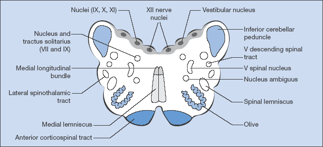
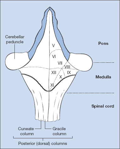
Remember diagrams of cross-sections of the brainstem and of the floor of the fourth ventricle (Figs. 6.2, 6.3) because these may greatly improve the analysis of a cranial nerve lesion. The following paragraphs outline a system for examination of the cranial nerves that recognises the need to examine parts of the head and neck in a logical order rather than by following the order of the cranial nerves directly.
Sense of Smell
Ask the patient:
‘Has there been any recent change in your sense of smell?’
If yes:
- assess informally with easily available substances (coffee granules, oranges)
- test formally with fresh ‘smell bottles’
Eyes
Observe and examine:
- visual acuity, either quickly with available literature, formally with Snellen charts or by counting fingers if vision is very poor
- visual fields to confrontation
 compare the patient’s visual fields with your own
 the patient’s head should be level with yours and at arm’s length away
 when testing the right eye, ask the patient to look straight into your left eye and vice versa for the patient’s left eye.
Ask the patient to:
‘Keep looking at my eye and tell me when you first see my finger out of the corner of your eye.’
- preferably using a red-headed hatpin, or your finger, bring the target towards the centre of the field of vision from the four diagonals (upper right, upper left, lower right, lower left)
- the nasal and superior fields are limited by the nose and eyebrow, respectively
- move the pin across the centre of each eye to examine for a central scotoma
- test for visual inattention (extinction and hemi-neglect) by asking the patient to identify which fingers you are moving with your hands held at the outer edges of the patient’s visual fields.
Ptosis
- third nerve lesion (complete ptosis)
- sympathetic lesion (partial ptosis) or as part of Horner syndrome
- generalised muscle weakness (myasthenia gravis, dystrophia myotonica, facio-scapulohumeral dystrophy, congenital lesions)
Pupillary Reflexes and Accommodation
- examine the pupillary reflexes in subdued light
- inspect the pupil for irregularity
- flash a pen-torch twice at each eye, once for direct and once for consensual responses; move the torch in from the side so as to avoid an accommodation–convergence reflex
- ask the patient to focus on your finger held at arm’s length and then move your finger to the patient’s focal point, observing convergence and pupillary constriction
External Ocular Movements (3rd, 4th and 6th Nerve)
Ask the patient:
‘Do you have any double vision?’
If the patient has not noticed diplopia:
- test the eye movements formally by asking the patient to follow your finger with their eyes, moving your finger slowly through the gaze regions
- examine upwards and downwards gaze for each eye in full abduction and adduction
- note any nystagmus.
Diplopia is maximal when looking in the direction of action of the paralysed muscle. The image further from the midline arises from the paralysed eye.
If the patient has noticed diplopia:
- ask in which direction it is worst
- move your forefinger in that direction and then ask if the two fingers that the patient sees are parallel to each other (lateral rectus palsy: 6th nerve) or at an angle (superior oblique palsy: 4th nerve)
- cover up each eye in turn and ask which image has disappeared.
Nystagmus
Ask the patient:
‘Look at my finger’
- hold the patient’s gaze in full abduction in each direction for a few seconds
- examine for nystagmus
- repeat with the eyes in upgaze.
Perform Fundoscopy
Ask the patient to:
‘Focus on a point at a distance ahead and slightly upwards’
Using the ophthalmoscope:
- check the red reflex
- focus through the lens and vitreous
- assess the optic disc, noting colour, cupping, margins and vessels
- examine the retina in all four quadrants
- observe the macula and major vessels.
Face
Facial Expression (7th nerve, Motor)
Ask the patient to:
‘Screw up your eyes very tightly’
- compare how deeply the eyelashes are buried on the two sides
‘Raise your eyebrows’
- compare right and left sides
‘Show your teeth’
- compare the nasolabial grooves.
Facial Sensation (5th Nerve, Sensory)
Test the three divisions on both sides:
- light touch with cotton wool
- corneal reflexes.
Mouth
Ask the patient to:
‘Clench your teeth’ (masseters, 5th nerve, motor)
- palpate the masseters
- test the jaw jerk: place one finger horizontally across the front of the jaw and tap the finger with a tendon hammer with the jaw relaxed and the mouth just open
‘Open your mouth and keep it open’ (pterygoids, 5th nerve, motor)
- attempt to force it closed
- in a unilateral lesion, the jaw deviates towards the weaker side
‘Say aaah’ (9th and 10th nerves: both mixed)
- observe the movement of the uvula and soft palate: normally they move upwards and remain central and the posterior pharyngeal wall moves little
- in a unilateral lesion the soft palate is pulled away from the weaker side
- test the gag reflex (9th nerve sensory, 10th nerve motor)
‘Put your tongue out’ (12th nerve)
Look for:
- wasting
- fasciculation
- in a unilateral lesion protrusion towards the weaker side.
Neck (11th nerve)
Observe:
- muscle wasting
Ask the patient to:
‘Lift your head off the pillows’
‘Put your chin on your right (or left) shoulder’
- resist the movement
- observe and palpate the sternomastoids
‘Shrug your shoulders’
- attempt to push them down
- observe and palpate the bulk of trapezius.
Hearing and Vestibular Function (8th Nerve)
Test hearing:
- whisper a number in each ear in turn, obstructing the contralateral ear
- perform Weber and Rinne tests (see below)
- inspect eardrums with an auriscope if indicated.
Test vestibular function if indicated:
- perform the Hallpike manoeuvre
- request caloric tests.
The Limbs
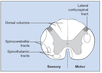
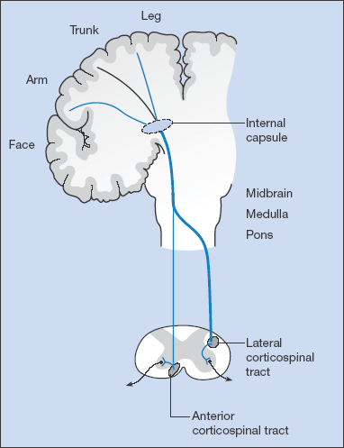
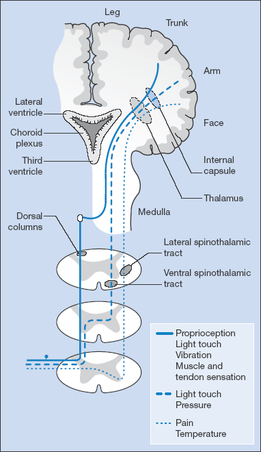
Figs. 6.4 and 6.5 give an overview of the motor and sensory systems.
Arms: Motor System
Observe:
- obvious muscle wasting
- fasciculation or tremor
- wasting of the small muscles of the hand, noting whether ulnar or thenar.
Test muscle tone:
- holding the arm, ask the patient to relax and move the arm gently at the elbow and wrist using an irregular rhythm to discourage resistance. Ask the patient to relax and, holding the thigh, shake the leg gently
- note cogwheel rigidity, which may be more obvious at the wrist.
Test muscle power in groups. Explain what you are doing to the patient:
‘I am going to test the strength of some of your muscles’
Shoulder (C5):
‘Hold both arms out in front of you and close your eyes’
Observe drifting of one arm indicating:
- weakness of the muscles at the shoulder
- loss of position sense with no evidence of weakness
- lesions of the cerebral cortex (when the patient will not be aware of the drift, sometimes even with open eyes).
Shoulder abduction:
‘Bend your elbows and lift your arms up to the side; don’t let me push them down’
- resist the action
Shoulder adduction:
‘Now pull them in towards your chest; don’t let me stop you’
- resist the action
Observe:
- winging of the scapula (nerve to serratus anterior, C5, 6, 7).
Elbow:
Flexion: C5, 6, 7 (biceps):
‘Bend your elbow, bring your hand to your shoulder; don’t let me straighten it’
Extension: C7: (triceps):
‘Now straighten your elbows and push me away’
Wrist:
Extension: C7:
‘Hold your arms out straight, cock up your wrists; don’t let me straighten them’
Hand grip: C8, T1:
‘Squeeze my fingers hard and stop me pulling them out of your grip’
Finger abduction: ulnar nerve:
‘Spread your fingers apart; don’t let me push them together’
- push against 1st and 5th fingers
Finger adduction: ulnar nerve:
Place a piece of paper between straight fingers
‘Don’t let me pull it out’
- attempt to pull the paper from between the patient’s fingers
Abduction of thumb: median nerve:
‘Place your hand down flat with the palm upward and the thumb overlying the forefinger. Lift the thumb vertically and don’t let me push it down’
Opposition of thumb: median nerve:
‘Put your thumb and little finger together and stop me pulling them apart with your forefinger’
Reflexes (Box 6.1)
 Box 6.1 A simple aide-mémoire for Reflexes
Stay updated, free articles. Join our Telegram channel

Full access? Get Clinical Tree


