Neuroendocrine Tumors of Gallbladder

 Well-differentiated neuroendocrine neoplasm (WDNEN): Grades 1 and 2


image Poorly differentiated neuroendocrine carcinoma (PDNC): Grade 3

– Small cell carcinoma, large cell neuroendocrine carcinoma


Clinical Issues


• WDNEN: All tumors have potential to metastasize

• PDNC: Dismal prognosis


Microscopic


• WDNEN
image Uniform round nuclei with minimal nuclear atypia

image Finely stippled (“salt and pepper”) chromatin

image Variable mitotic activity, but < 20 per 10 HPF

image No or minimal necrosis

• Small cell carcinoma
image Small, round or fusiform hyperchromatic nuclei

image Finely dispersed granular chromatin

image Nuclear moulding

image High mitotic rate

image Often show extensive necrosis &/or frequent apoptotic bodies

• Large cell neuroendocrine carcinoma
image Large polygonal cells

image Nucleoli are often present and may be prominent

image Variable amounts of cytoplasm but usually abundant

image Frequent necrosis

• Grading by mitotic rate and Ki-67 labeling index
image Grade 1 (low grade): Mitosis < 2 per 10 HPF and Ki-67 < 3%

image Grade 2 (intermediate grade): Mitosis 2-20 per 10 HPF &/or Ki-67 3-20%

image Grade 3 (high grade): Mitosis > 20 per 10 HPF &/or Ki-67 > 20%

image
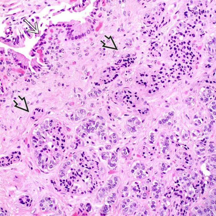
Incidental Well-Differentiated Neuroendocrine Neoplasm
This 3-mm, low-grade well-differentiated neuroendocrine neoplasm (WDNEN) (carcinoid) image was incidentally found at the gallbladder neck. Cholecystectomy was performed for cholelithiasis. The overlying gallbladder mucosa image appears unremarkable.


image
Nested Growth Pattern
Tumor cells of this gallbladder WDNEN show diffuse cytoplasmic immunoreactivity to antichromogranin antibody, which highlights a nested growth pattern. Same staining results are also observed for synaptophysin.

image
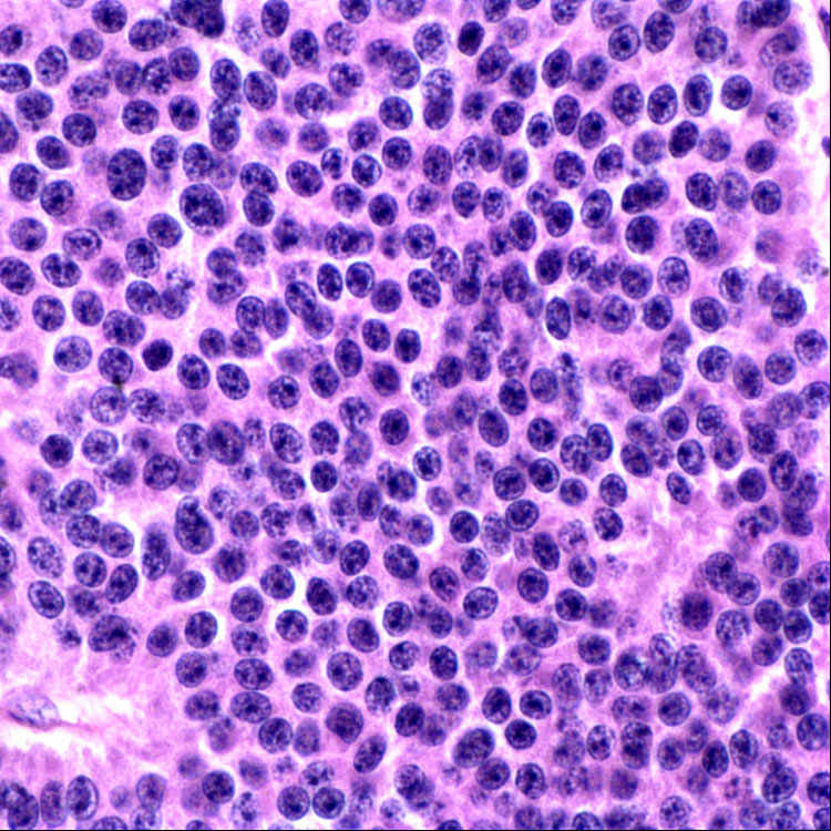
Histologic Features
Tumor cells of WDNEN typically have uniform round nuclei, finely stippled (“salt and pepper”) chromatin, inconspicuous nucleoli, and eosinophilic cytoplasm. No mitotic figures or necrosis are seen in this microphotograph.

image
Clear Cell Variant
This WDNEN shows nested tumor cells, the majority of which have abundant foamy cytoplasm due to lipid accumulation image
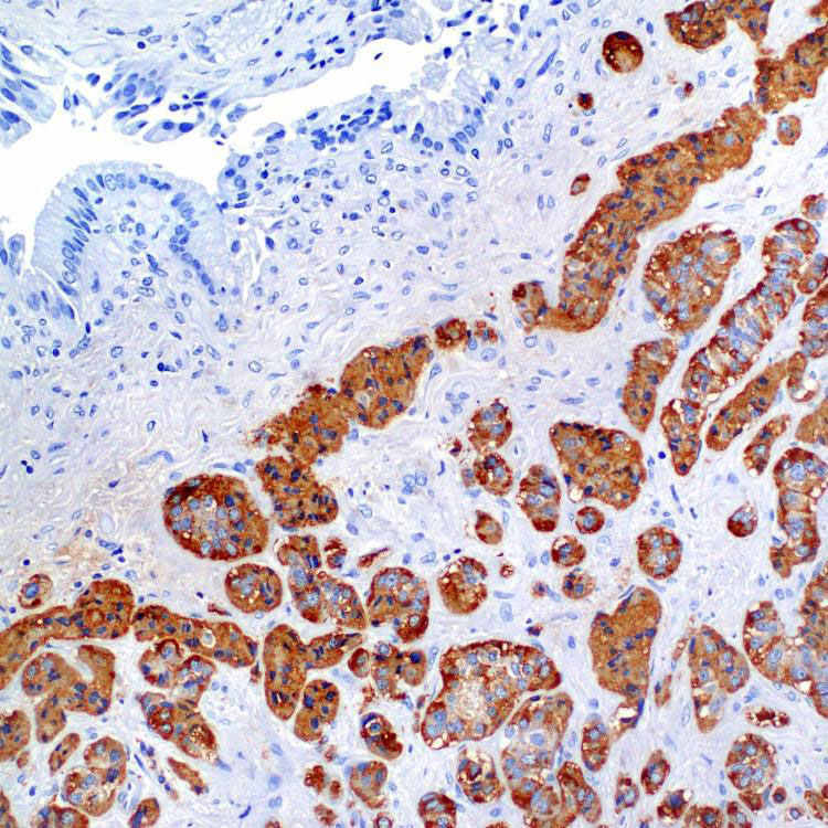
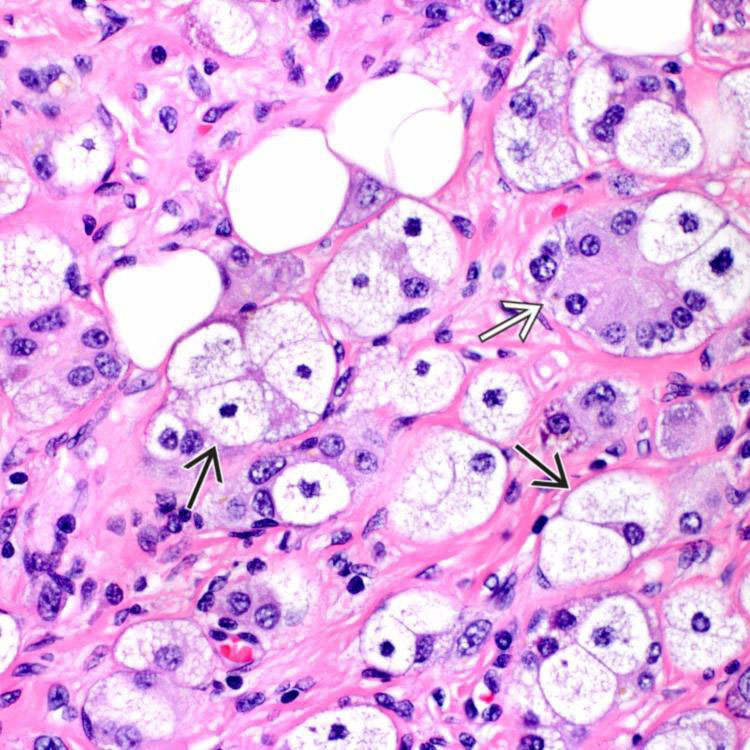
. Tumor cells with eosinophilic cytoplasm are also present image .


TERMINOLOGY


Abbreviations


• Well-differentiated neuroendocrine neoplasm (WDNEN)

• Poorly differentiated neuroendocrine carcinoma (PDNC)


Synonyms


• WDNEN
image Well-differentiated neuroendocrine tumor

image Carcinoid tumor (not preferred)

• PDNC
image Small cell carcinoma
– Small cell neuroendocrine carcinoma

– Small cell undifferentiated carcinoma

– Oat cell carcinoma

image Large cell neuroendocrine carcinoma

image High-grade neuroendocrine carcinoma


Definitions


• Neoplasms with neuroendocrine differentiation arising in gallbladder, including WDNEN and PDNC


ETIOLOGY/PATHOGENESIS


Disease Association


• PDNC is frequently associated with gallstones

• WDNEN is infrequently associated with von Hippel-Lindau disease and multiple endocrine neoplasia type 1


CLINICAL ISSUES


Epidemiology


• Incidence
image 0.2-0.3 per 100,000 population

– WDNEN: 0.2% of all sites

– PDNC: 4.0% of all gallbladder malignancies

image Small cell carcinoma > large cell

• Age
image Older adults, usually 6th-7th decades of life
– Range: 25-85 years

• Sex
image Slightly more common in females


Presentation


• WDNEN: Usually asymptomatic
image Incidental finding after cholecystectomy for cholelithiasis or cholecystitis in most cases

image Abdominal pain in occasional cases

image Not typically associated with carcinoid syndrome

Only gold members can continue reading. Log In or Register to continue

Stay updated, free articles. Join our Telegram channel

Apr 20, 2017 | Posted by in PATHOLOGY & LABORATORY MEDICINE | Comments Off on Neuroendocrine Tumors of Gallbladder

Full access? Get Clinical Tree

Get Clinical Tree app for offline access