Myositis Ossificans
Elizabeth A. Montgomery, MD
Key Facts
Clinical Issues
Mean age: ˜ 30 years (range: 1-95 years)
M > F (˜ 3:2) for classic lesions
F > M for digital lesions
Microscopic Pathology
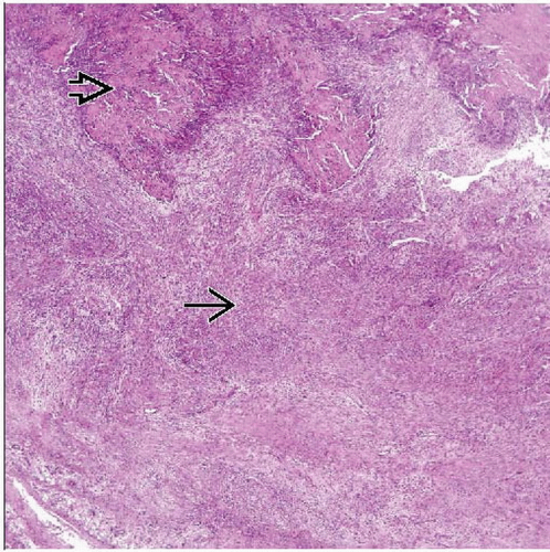
Zonated appearance with cellular central area with peripheral ossification
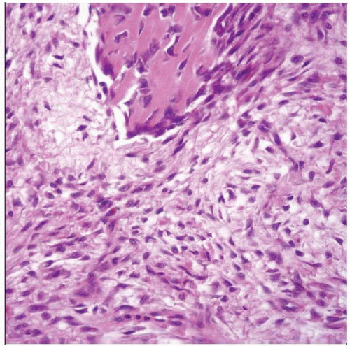
Cellular central portion composed of short fascicles of myofibroblasts
Numerous mitoses (not atypical)
Vascular stroma with myxoid areas, fibrin, extravasated erythrocytes
Peripheral portion with bone formation
Trabeculae of woven bone rimmed by osteoblasts
Older lesions show mature-appearing bone
Digital lesions lack zonation
Cartilage uncommon but occasionally encountered
TERMINOLOGY
Synonyms
Fibroosseous pseudotumor of digits (for finger lesions)
Pseudomalignant osseous tumor of soft tissue
Definitions
Localized self-limited benign lesions composed of cellular myofibroblastic proliferation with ossification
CLINICAL ISSUES
Epidemiology
Incidence
Rare
Age
Mean age: ˜ 30 years
Range: 1-95 years
Gender
M > F (˜ 3:2) for classic lesions
F > M for digital lesions
Presentation
Arise in areas prone to local trauma
Elbow, thigh, buttock, shoulder
Rapidly growing, variably tender mass


