Continuum with lesions termed myopericytoma and infantile hemangiopericytoma
Clinical Issues
• Most common from birth to 2 years
• Most solitary examples in subcutaneous tissues of head and neck
• Simple excision for solitary lesions
• Outcome for multicentric form is function of involved sites
Extensive lung involvement poor prognostic factor
Microscopic
• Biphasic pattern classic
• Myoid nodules separated by cellular pockets with hemangiopericytoma-like vascular pattern
• Variable amounts of each component may be present
• Most cases have minimal atypia and mitotic activity
• Spindle cell areas
Often prominent beneath ulcerated mucosal surfaces
Ancillary Tests
• Usually label with α-actin and calponin, but negative to focal desmin, caldesmon
• Negative for S100 and cytokeratins
• No characteristic alterations or mutations in myofibroma
• Mutations detected in PDGFRB, NOTCH3, NDRG4 in myofibromatosis
Top Differential Diagnoses
• Smooth muscle tumors
• Fibromatosis
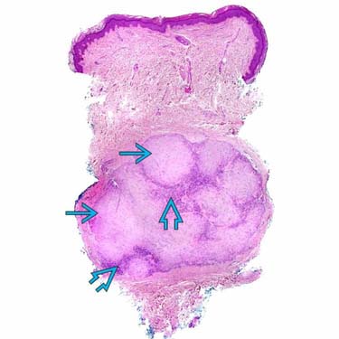
Myofibroma at Scanning Magnification This punch biopsy shows a deep-seated dermal tumor. Note the prominent lobulations and a biphasic pattern, consisting of myoid nodules and intervening hemangiopericytomatous zones.
Myofibroma With Hemangiopericytomatous Areas This example of cutaneous myofibroma has prominent hemangiopericytomatous zones .
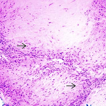
Myofibroma, Higher Magnification of Myoid Lobules Higher magnification shows the myoid lobules separated by more cellular areas. The myoid cells show cytoplasmic eosinophilia .
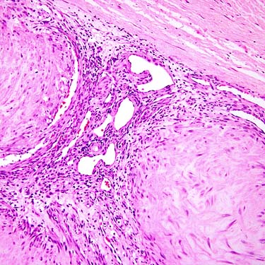
Myofibroma, Hemangiopericytomatous Zone Higher magnification shows the hemangiopericytoma-like component of a myofibroma.


 and intervening hemangiopericytomatous
  zones.
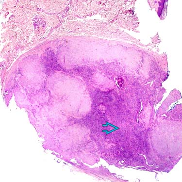
 .
.
 .
.