Muscle Biopsy in Neuromuscular Diseases


Muscle Biopsy in Neuromuscular Diseases


Reid R. Heffner Jr.

Steven A. Moore

Lucia L. Balos


This chapter is designed to provide the surgical pathologist with a concise discussion of commonly encountered nonneoplastic neuromuscular diseases. Like all of medicine, surgical pathology is changing and becoming more specialized, such that in today’s world, many pathologists will not choose to render a final diagnosis in a muscle biopsy. Many cases will be referred to an expert at a center with a muscle pathology laboratory. Depending on the proximity of the specialized laboratory, these cases may be sent out at any stage from fresh tissue on wet ice to unprocessed frozen and fixed tissues to a partially worked-up case consisting of glass slides with or without frozen tissue. Our approach is to focus on morphology and on those disorders amenable to diagnosis by commonly available morphologic techniques. However, we include in many instances other information such as molecular testing for the pathologist’s consideration. Readers who wish to acquire a more detailed understanding of the field are referred to a variety of textbook and website resources (1,2,3,4,5,6,7,8,9,10,11,12 and 13).


CLINICAL INFORMATION

Clinical information is critical to structuring the evaluation of a given muscle biopsy, and clinicopathologic correlation is essential for accurate interpretation. The ordering physician is asked to provide pertinent clinical data on a requisition that is not so lengthy as to discourage compliance. Personal communication is encouraged as it is almost as useful as the microscope in interpreting some muscle biopsies.


COLLECTION AND PREPARATION OF THE MUSCLE BIOPSY SPECIMEN

Ideally, muscle biopsies should be performed by persons skilled in biopsy technique and with knowledge of specimen submission procedures. The requesting physician, who is familiar with the patient, is obligated to ensure that an appropriate muscle is sampled. The biopsy specimen should be representative of the disease process. For example, if the symptoms involve the legs rather than the arms, a specimen from the upper extremity cannot be expected to reflect the disease process accurately. Moreover, the specimen should be obtained from a muscle in which the disease process is active and evolving. A specimen from a severely affected muscle, in which marked weakness or atrophy is present, is generally characterized by end-stage histopathology that is difficult to interpret, much like end-stage renal disease. Furthermore, the biopsy should be obtained from the belly of the muscle and deep to the fascia, unless fasciitis is being considered in the differential. This will reduce the likelihood of including myotendinous junctions that can be confused with myopathic changes. Sites of needle sticks (e.g., electromyography [EMG] or immunization) or other trauma should be avoided.

An accurate pathologic diagnosis depends on obtaining biopsy specimens of adequate size and quality followed by proper laboratory processing of the muscle sample. Because of the unique requirements for handling the muscle specimen, the biopsy must be done at a time when experienced technical assistance is available. Before excision from the patient, the muscle specimen can be maintained in an isometric state by using a muscle clamp, which limits the contraction artifact caused by cutting the muscle. Because the muscle is placed in the instrument lengthwise, the specimen is already oriented for processing. Inasmuch as the biopsy specimen must extend entirely across the clamp, an acceptable sample size of approximately 0.5 cm in diameter and 1 cm in length is more likely to be provided. The specimen should be of sufficient size to permit the adequate observation of the pathologic process. At least two separate specimens are routinely requested. Biopsies should be transported to the laboratory as fresh tissue wrapped in saline-moistened gauze. Too much saline, especially immersion in saline, is to be avoided, as it introduces artifacts when the muscle is frozen.

Once in the laboratory, muscle tissue is distributed (triaged) to best evaluate the pathology. Clinical context and the amount of tissue received may drive decisions. However, a standard approach is to distribute tissue to facilitate each of the following: cryosection histology, epon embedment for possible electron microscopy, stored frozen tissue for possible biochemical or molecular studies, and paraffin embedment for standard histology (see Fig. 4.1). The biopsy specimen with the best gross appearance is typically used to first trim a thin strip for fixation in glutaraldehyde, then orient and freeze the remainder for cryosections. Additional biopsy specimens are divided between storage as frozen tissue and fixation in formalin for the generation of paraffin blocks.

The portion of the biopsy chosen for cryosections should optimally be about 0.5 cm in diameter and 1.0 cm in length. A variety of flash-freezing techniques have been described (14,15). One commonly used method is to orient the muscle for eventual cross-sectional histology (see Fig. 4.1), then immerse the biopsy in isopentane precooled to —160°C. All frozen tissue should be stored wrapped in foil inside an airtight container (e.g., polycon). Most laboratories use a —80°C freezer for specimen storage.


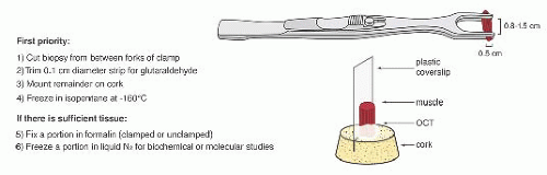
FIGURE 4.1 Muscle biopsy in a clamp with suggested priorities for processing a routine skeletal muscle biopsy specimen. OCT, optimal cutting temperature embedding medium.


A standard histologic approach is to produce a hematoxylin and eosin (H&E) slide from each paraffin block and to cut serial frozen sections for H&E, Gomori trichrome, fiber typing by either adenosine triphosphatase (ATPase) or myosin heavy chain immunostaining, and nicotinamide adenine dinucleotide, reduced (NADH-TR). Cryosections for these standard stains are typically cut at a thickness of 10 µm. When needed, a wide range of histochemical, enzyme histochemical, immunoperoxidase, and immunofluorescence techniques can be applied to paraffin or frozen sections. Epon sections stained with toluidine blue with or without subsequent electron microscopy provide additional insight in selected cases. In a small subset of cases, ultrastructural features can be critical to reaching a specific diagnosis.


INTERPRETATION OF THE MUSCLE BIOPSY SPECIMEN


NORMAL MUSCLE

In the evaluation of the muscle biopsy specimen, familiarity with the normal structure is the basis for understanding the diversity of pathologic reactions that occur in neuromuscular disease. The reader is referred elsewhere for a more detailed discussion of the light microscopy, histochemistry, and electron microscopy of normal muscle (2,3 and 4). The myocyte is a multinucleated, syncytium-like cell with a shape resembling an elongated cylinder, although the normal adult fiber is not truly round but, instead, is polygonal or multifaceted in cross section (Fig. 4.2). The sarcolemmal nuclei are ordinarily located peripherally, often more than one per fiber in transverse sections. The diameter of the fibers depends on several factors. In general, powerful proximal muscles are made up of fibers with a mean diameter (50 to 70 µm) greater than that of smaller, distal, or ocular muscles (20 µm), which are devoted to finely coordinated activity. Fiber size is greater in males than in females, probably because of hormonal influences and the general propensity of males to engage in more strenuous physical activity. That exercise encourages fiber hypertrophy in both sexes is well established. Muscle fibers in children and the elderly are smaller than those in young, healthy adults.

In vertebrates, notably certain species of birds, one can distinguish between red (e.g., soleus) and white (e.g., pectoralis) muscles, with the color of red muscles being a consequence of their greater myoglobin content. Red muscle, with its larger mitochondrial population and higher capillary density, is adapted to aerobic respiration and is designed for postural or sustained activity. White muscle, which contains fewer mitochondria and abundant glycogen, is capable of anaerobic respiration; it is more suited to sudden and intermittent action. Although an entire muscle in lower animals may be composed of either red or white fibers, human muscle is constructed of both fiber types, which are arranged in a mixed mosaic pattern resembling a checkerboard. The location of the muscle in the body and its function determine the proportion of type 1 and type 2 fibers, but the average muscle has about twice as many type 2 fibers (60% to 65%) as type 1 fibers (35% to 40%) (16).

Fiber typing, the demonstration of the histochemical properties of muscle fibers within a biopsy specimen, is accomplished by carrying out enzyme histochemistry in frozen sections (Table 4.1). Fiber types are not evident in slides stained with H&E. The traditional approach is to perform myofibrillar ATPase reactions at acidic and alkaline pH. In the standard or alkaline ATPase reaction conducted at a pH of 9.4, type 1 fibers appear light (they can be seen better in sections counterstained with eosin) and type 2 fibers appear dark (Fig. 4.3). Fibers with intermediate staining properties are not seen in the alkaline reaction.
When the pH of the incubating solution is reduced to the acidic range (pH 4.2) in the so-called reverse ATPase reaction, type 1 fibers are very dark and type 2 fibers are very light. At a slightly less acidic pH 4.6, type 2A fibers are very light and the staining intensity of type 2B fibers is intermediate. Commercial antibodies to slow (type 1) and fast (type 2) myosin are now available. By means of automated immunoperoxidase staining, fiber typing with results similar to those obtained with ATPase reactions can be achieved in either cryosections or paraffin sections (3,17).


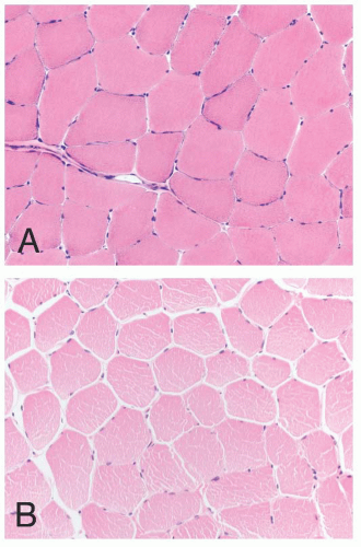
FIGURE 4.2 Normal muscle in transverse section (A, frozen; B, paraffin embedded). The fibers are typically polygonal, and the nuclei are located peripherally. Empty space between muscle fibers is a shrinkage artifact, which is typically more pronounced in paraffin sections (H&E).


TABLE 4.1 Fiber Typing in Skeletal Muscle


@


Type 1


Type 2


Gross color


Red


White


ATPase activity at pH 9.4


Low


High


Myosin heavy chain


Slow


Fast


Oxidative enzyme content


High


Low


Glycogen content


Low


High


Phosphorylase activity


Low


High


Lipid content


High


Low


ATPase, adenosine triphosphatase.


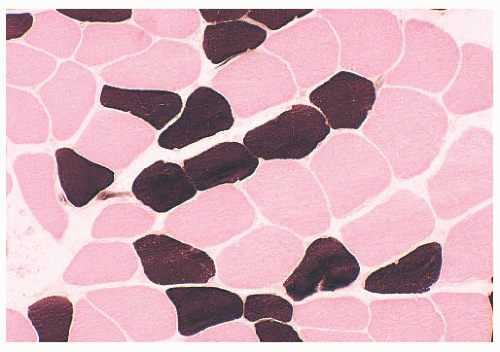
FIGURE 4.3 Fiber types in normal muscle. In the alkaline adenosine triphosphatase (ATPase) reaction, type 1 fibers are light and type 2 fibers are dark (ATPase at pH 9.4 and counterstained with eosin).

The oxidative stains, such as the NADH-TR reaction or specific mitochondrial enzyme histochemistry (e.g., succinic dehydrogenase or cytochrome C oxidase), reflect the concentration of mitochondria within the myofibers. The fiber staining is essentially the opposite of that seen in the alkaline ATPase reaction. Thus, intensely stained fibers are designated as type 1 (oxidative), and lighter fibers are designated as type 2. Typically, oxidative enzyme reactions further divide type 2 fibers into two subpopulations. Type 2B fibers are very lightly stained, whereas the staining intensity of type 2A fibers is intermediate between that of type 1 and that of 2B. The myofibril network is stained such that the sarcoplasm has a regular, fine granular appearance.

All muscle fibers contain phosphorylase as well as glycogen; these are more abundant in type 2 (glycolytic) fibers. Some laboratories take advantage of the histochemical reaction for phosphorylase, or the periodic acid-Schiff (PAS) stain, as a means of fiber typing, but our experience dictates that such staining is unpredictable, especially because much of the glycogen leaches out of the muscle sections during processing. We restrict use of the phosphorylase reaction to cases of possible enzyme deficiency (McArdle disease). Lipid vacuoles are more numerous in the sarcoplasm of type 1 fibers, yet fat stains such as oil red O seldom demonstrate an unequivocal difference between fiber types. Fat stains are more valuable when the diagnosis of lipid storage myopathy is suspected.


ARTIFACTS

Among the most unavoidable and disturbing artifacts is the vacuolation produced by ice crystals that form during improper freezing of the muscle biopsy specimen (Fig. 4.4). It may also occur after improper transport or storage that allows thawing and refreezing of the sample. This type of artifact may simulate pathologic change, such as vacuolar myopathy, or it may distort pathologically altered fibers, thereby precluding proper interpretation. In contrast to pathologic vacuoles, the vacuoles associated with ice crystals are in the more slowly frozen or earlier thawing portion of the specimen, which is generally in the center or at the periphery of the tissue block, respectively. Vacuoles tend to affect every fiber in the region of artifact and may be arranged in a gradient according to size. Where the artifact is minimal, small vacuoles are numerous in each fiber, uniform in size, and arranged in a waffle pattern; they are located between the myofibrils. In poorly frozen regions, larger clear vacuoles are present. Muscle transported to the laboratory in saline can acquire artifactual separation of muscle fibers during freezing due to the expansion of water that accumulated in the interstitium.


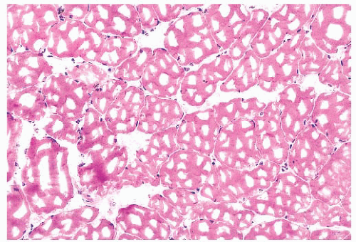
FIGURE 4.4 Freezing artifact. Extensive vacuolar change can be caused by the formation of ice crystals in the muscle during improper freezing. Vacuoles can be a variety of geometric shapes (H&E).

With severe contraction artifact in longitudinal sections, dark contraction bands alternate with lucent, disrupted zones within the fiber (Fig. 4.5). The latter appear as jagged cracks in the sarcoplasm in transversely oriented sections. Contraction artifact is most noticeable at the edges of the specimen. This artifact is commonly seen in unclamped specimens, when
plastic disposable clamps that only partially prevent contraction are used or when the muscle is injected with local anesthetic. Contraction artifact renders muscle particularly unfit for electron microscopy. The orderly structural landmarks are obliterated by myofibrillar fragmentation and disorientation.


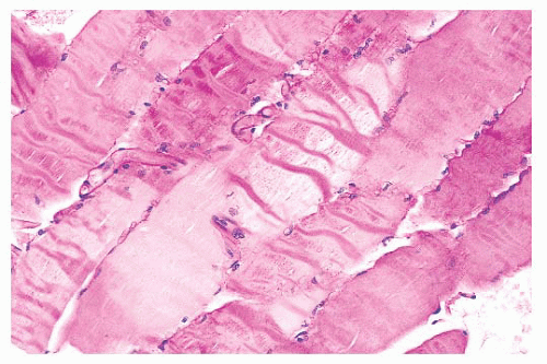
FIGURE 4.5 Contraction artifact. Darker contraction bands and disrupted lucent zones are seen in several longitudinally oriented fibers (PAS).


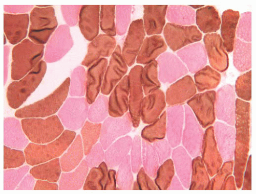
FIGURE 4.6 Muscle fiber wrinkling. Frozen sections may partially lift off the slide during staining, creating artifacts such as stripes and ring structures in the fibers (ATPase, pH 9.4, counterstained with eosin).

A frequent artifact occurs in frozen sections when the section lifts partially off the slide. The staining intensity of the fibers varies, and a variety of striped or ring structures result from curling or wrinkling of the fibers (Fig. 4.6).


GENERAL PATHOLOGIC REACTIONS OF MUSCLE

When evaluating muscle biopsies, the surgical pathologist is likely to come across one or more of the features described in the next few paragraphs. Very few of the pathologic changes that may be observed in the muscle biopsy specimen are totally specific for a single disease, although each has diagnostic significance because it is restricted to one disease or a limited number of diseases. The presence of each pathologic change described in the following sections should engender a differential diagnosis in the mind of the observer. Arrival at a precise diagnosis involves correlation not only with the clinical information and laboratory test results but also integrating the various pathologic findings to arrive at a pattern or signature of the disease process. All of the pathologic changes discussed here are light microscopic features, and only some are appreciated in greater detail ultrastructurally. Where appropriate, a description or illustration of the fine structure accompanies the description of the light microscopic features.


Variations in Muscle Fiber Size: Smallness, Atrophy, and Hypertrophy

Undoubtedly, the most common abnormality encountered in muscle biopsy pathology is variation in fiber size. Diameters range widely with the age, sex, and physical activity of the patient. At birth, muscle fiber diameters average 10 to 15 µm, whereas in adults, the normal range is from 40 to 80 µm (3). One of the most demanding challenges for the surgical pathologist is interpretation of hypertrophy and abnormal smallness due to developmental abnormalities or atrophy when abnormal fiber size is the predominant pathologic finding. The usefulness of histochemistry can be considerable in this situation. Because the muscle fiber depends on neural and other influences for survival, the disruption of these influences results in the atrophy of the fiber. The most common form of atrophy in neuromuscular disease is that caused by denervation. In addition, the maintenance of muscle fiber integrity requires regular activity. Disuse, as may occur with prolonged bed rest, can lead to muscle atrophy. Finally, a reduction in fiber volume may be a complication of aging, ischemia, or poor nutrition.


TABLE 4.2 Diseases with Prominent Type 1 Fiber Atrophy/Smallness


Myotonic dystrophy


Nemaline myopathy


Centronuclear myopathy


Congenital fiber-type disproportion


Hypertrophy, by comparison, is principally a consequence of an increase in muscular work, either during exercise or as a compensatory reaction of normal, intact fibers to the atrophy of others in their midst. In an evaluation of fiber size, the measurement of cell diameters may be fruitful. A quick evaluation of the range of diameters can be performed using the pointer arrow after measuring the arrow at various magnifications with a glass slide or eyepiece micrometer. Morphometric analysis of the muscle specimen may be indicated in the event that changes in fiber diameters are minimal and subtle. Morphometry can be performed manually with an eyepiece micrometer or electronically with a computer-assisted image analyzer (18). To obtain statistically significant morphometric data, the lesser diameter of each muscle fiber should be measured, and the minimum number of fibers in the sample should be 200. Histograms of each fiber type can be generated from ATPase- or myosin heavy chain immunoperoxidase-stained sections.

The atrophic or hypertrophic process may be selective, involving only one fiber type, or nonselective (19). Selective atrophy of type 1 fibers is seen most commonly in myotonic dystrophy but is also seen in distal myopathy, nemaline myopathy, centronuclear myopathy, and congenital fiber-type disproportion (Table 4.2). Type 2 fiber atrophy (Fig. 4.7) is observed
in myasthenia gravis, acute denervation, disuse, and systemic malignancy (20). In our experience, more than half of such cases are attributable to corticosteroid therapy (Table 4.3). Hypertrophy restricted to type 1 fibers is relatively specific for infantile spinal muscular atrophy (SMAI), although such hypertrophy may develop in normal athletes who undergo endurance training. Type 2 fiber hypertrophy has been reported in runners, notably sprinters, and in congenital fibertype disproportion. Non-fiber type-selective alterations in fiber size are somewhat uninformative from a diagnostic standpoint. Denervation accounts for a large proportion of cases of nonselective atrophy but certainly not all of them. Hypertrophy involving both major fiber types is frequently noted in muscular dystrophy, inclusion body myositis, myotonia congenita, and acromegaly.


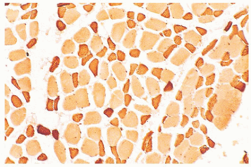
FIGURE 4.7 Type 2 fiber atrophy in a patient with malignancy and cachexia (immunostain for fast myosin).


TABLE 4.3 Diseases with Prominent Type 2 Fiber Atrophy


Corticosteroid therapy and hypercorticoidism


Myasthenia gravis


Disuse atrophy


Acute denervation


Paraneoplastic myopathy


The pattern of atrophy may be diagnostic in some atrophic diseases. Grouped atrophy, which is recognized as a clustering of five or more small angular fibers, is essentially pathognomonic for chronic neurogenic disorders (Fig. 4.8). Panfascicular atrophy, an extreme version of grouped atrophy wherein the vast majority of fibers in each fascicle are severely atrophic, is a distinctive feature of SMAI. Perifascicular atrophy typifies dermatomyositis, in which fiber atrophy is limited mainly to the periphery of the fascicles. Unfortunately, in many muscle biopsy specimens, atrophic fibers are randomly situated in the section. This pattern of atrophy is totally nonspecific.


Nuclei

One of the more frequent and easily recognized abnormalities observed in muscle biopsies is the internalization of muscle fiber nuclei. According to quantitative studies, the nuclei in cross sections of normal muscle are peripheral (subsarcolemmal) in 97% to 99% of fibers. In specimens from patients with neuromuscular disease, the counts of internal nuclei are commonly elevated to 5% to 10% of fibers. Although the sarcoplasmic integrity appears undisturbed, such fibers are often mildly or moderately altered in size (Fig. 4.9). This inconstancy of nuclear position is of no specific diagnostic import, and it is a reaction to a variety of diverse injuries. However, if most of the fibers contain randomly distributed internal nuclei, the diagnosis is more likely a myopathic condition (Table 4.4). The virtually pathognomonic criterion of centronuclear myopathy is the presence of a single central or paracentral nucleus within most muscle fibers. As opposed to nuclear internalization in the absence of injury to the sarcoplasm, after myonecrosis, the nuclei may no longer be subsarcolemmal. They commonly migrate internally in degenerating and regenerating fibers, no matter what the cause. Severely atrophic fibers also contain multiple nuclei that seem to remain intact, forming pyknotic nuclear clusters as the sarcoplasm is progressively diminished in volume. Most often seen in neuropathic disorders or end-stage muscle, these nuclear clusters can be mistaken for lymphocytic inflammation.


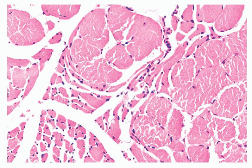
FIGURE 4.8 Chronic neurogenic atrophy. Angulated atrophic fibers arranged in groups are denervated motor units. The large size of the groups is suggestive of a chronic neuropathic process.


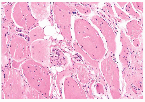
FIGURE 4.9 Internally placed nuclei. Many fibers contain one or more internal nuclei (H&E).


Fiber Splitting

Muscle fibers that have become hypertrophic with multiple internalized nuclei commonly split into smaller subunits of two or more smaller fibers that appear to be mature myocytes with intact sarcoplasm. Before splitting is concluded, the fiber assumes a segmented appearance as slitlike spaces form invaginations between individual segments (Fig. 4.10). Within
each space, the extensions of the plasma membrane remain continuous around the dividing portions of the cell. Fiber splitting is typically more conspicuous in chronic necrotizing myopathies (e.g., muscular dystrophies or inflammatory myopathies) (21). Internalization of capillaries sometimes accompanies the fiber splitting process (see panel A of Fig. 4.36).


TABLE 4.4 Conditions in Which Internal Nuclei Have Diagnostic Significance


Myotendinous insertion


Congenital myopathies, especially centronuclear myopathy


Muscular dystrophies, especially myotonic dystrophy


Other necrotizing myopathies


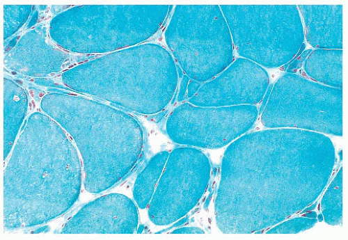
FIGURE 4.10 Fiber splitting. Several hypertrophic fibers are seen. The fiber at the bottom and center has been split into two smaller subunits (Gomori trichrome).


Fiber Shape

Assessing fiber shape is typically more accurate in frozen sections than in paraffin sections. In contrast to the normal polygonal contour of the myofiber viewed in the transverse plane, rounded fibers favor a myopathic process, particularly muscular dystrophy or congenital myopathy. In neuropathic diseases (excluding SMAI where small fibers are rounded), the atrophic fibers are angular (or ensiform) and grouped (Fig. 4.11). These fibers appear flattened and narrow with tapered or pointed ends. Grouping is the result of simultaneous atrophy of entire motor units. Angulated atrophic fibers are also found in myasthenia gravis, disuse, myotonic dystrophy, and steroid myopathy.


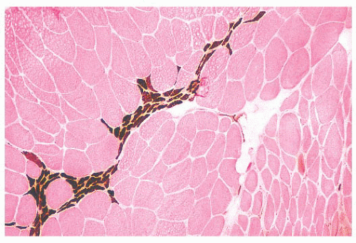
FIGURE 4.11 Fiber type grouping in denervation with reinnervation. Grouping of fiber types replaces the normal checkerboard staining pattern (ATPase at pH 9.4).


TABLE 4.5 Inflammation Seen in the Biopsy Specimen


Pathologic Features


Disease


Perivascular, angiocentric


DM, connective tissue disease


Endomysial, around fibers


PM, IBM, viral


Nodular


Rheumatoid arthritis, granulomas


Polymorphous with eosinophils


PAN, drug reactions, trichinosis, eosinophilic fasciitis


DM, dermatomyositis; IBM, inclusion body myositis; PAN, polyarteritis nodosa; PM, polymyositis.


Inflammation

Interstitial inflammatory infiltrates are most frequently encountered in immunologically mediated or idiopathic inflammatory myopathies (Table 4.5). Most important among these are polymyositis (PM), dermatomyositis (DM), and inclusion body myositis, which make up nearly a third of all cases evaluated at the University of Buffalo. The inflammatory cells in PM invade the endomysium, sometimes enveloping necrotic fibers. Sheets of inflammatory cells expand the endomysial spaces in acute, more severe disease (Fig. 4.12). In DM, they surround intramuscular blood vessels with minimal infiltration of the vascular walls. The inflammatory cells are mononuclear in type, chiefly consisting of mature lymphocytes. Plasma cells are a minor part of the inflammatory response. Rare eosinophils are seen, and neutrophils are absent.

An inflammatory myopathy histologically identical to PM and DM may accompany any of the systemic connective tissue diseases. However, crucial differences among several of these collagen vascular disorders are diagnostically reliable. Nodular infiltrates composed largely of plasma cells are highly suggestive of rheumatoid arthritis. Polyarteritis nodosa and systemic lupus erythematosus, on the other hand, are typically associated with a vasculitis. Polyarteritis nodosa shows an affinity for larger vessels, especially arteries, within the epimysium and perimysium. The entire wall of the vessel may be infiltrated by inflammatory cells, which usually include conspicuous eosinophils.
In systemic lupus erythematosus, the affected vessels are smaller in caliber, and they exhibit fibrinoid necrosis. Areas of vascular injury attract neutrophils, and the remnant of their breakdown, nuclear dust, is also seen. With the use of immunohistochemical techniques, deposits of immunoglobulin and complement can be demonstrated at sites of vascular injury in both conditions.


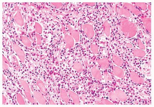
FIGURE 4.12 Inflammatory myopathy. Sheets of lymphocytes surround muscle fibers and expand the endomysium (H&E).

Granulomatous inflammation may be indicative of sarcoidosis or idiopathic granulomatous myositis, a chronic progressive myopathy of middle-aged women without systemic manifestations (22,23). The granulomas in both are sharply circumscribed and nonnecrotizing. They invade and expand the interstitium, and they are composed of lymphocytes, epithelioid histiocytes, and multinucleated giant cells. Trichinella infestation may elicit a granulomatous response in muscle that usually contains numerous eosinophils. Other microbial agents, including bacteria, fungi, and viruses, may cause inflammatory myopathy, but a muscle biopsy is not routinely a part of the diagnostic investigation of these infections.


Fiber Necrosis (Myonecrosis) and Regeneration

The initial sign of necrosis in light microscopic sections is an alteration in the tinctorial properties of the muscle fiber that is perhaps best appreciated in H&E stains. The acutely necrotic fiber first stains more intensely eosinophilic and then pales to a wan shade of pink. Simultaneously, there is a loss of striations in the sarcoplasm. The myocyte nuclei may become fragmented and eventually are no longer visible. As the process of myonecrosis proceeds, the sarcoplasm is evacuated by macrophages that eventually occupy the entire space within the original myocyte basement membrane (Fig. 4.13; also see Fig. 4.29 later in this chapter). Often, even before the total removal of the necrotic sarcoplasmic debris, the phase of regeneration has supervened; therefore, both regenerative and phagocytic activity may be seen in the same fiber. Necrosis may be a part of the pathologic response in many muscle diseases, but it is prevalent in muscular dystrophies (especially Duchenne and related limb-girdle muscular dystrophies) and the inflammatory myopathies (Table 4.6).

Hyaline fibers are pathologically rounded and enlarged. They are more deeply stained than normal fibers, whether in paraffin or frozen sections stained with H&E, trichrome, PAS, or histochemical methods (Fig. 4.14). The sarcoplasm is smudged and homogeneous. The hyaline fiber is a bona fide pathologic change in tissue unaffected by contraction artifact, and in many instances, it represents an early stage of cell necrosis (24). Serial sections through the same fiber may reveal zones of unequivocal necrosis and phagocytosis adjacent to hyalinization. Hyaline fibers are most commonly encountered in Duchenne muscular dystrophy, and they are most numerous in this condition.


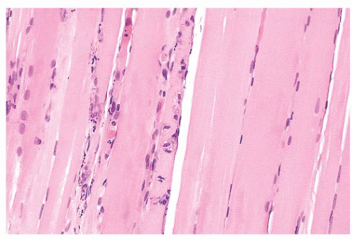
FIGURE 4.13 Fiber necrosis (myonecrosis). The necrotic process in the fiber at the center of this longitudinal section is recognized by a loss of cross-striations and early phagocytosis (H&E).


TABLE 4.6 Fiber Necrosis Seen in the Biopsy Specimen


Pathologic Features


Disease


Small groups of necrotic fibers


Muscular dystrophies, especially Duchenne and other dystrophinglycoprotein complex dystrophies


Perifascicular necrosis


Dermatomyositis


Random fiber necrosis


PM, IBM, muscular dystrophies


Infarcts with large areas of necrosis


PAN, compartment syndrome


Extensive, diffuse


Rhabdomyolysis in patients with CPT deficiency, alcoholics, military recruits


CPT, carnitine palmitoyltransferase; IBM, inclusion body myositis; PAN, polyarteritis nodosa; PM, polymyositis.


Fiber necrosis (myonecrosis) generally acts as a stimulus for subsequent regeneration. Hence, the presence of regenerating fibers in a biopsy specimen, even in the absence of necrotic fibers, is a likely indicator of previous necrosis. The regeneration of fibers is believed to arise primarily from the proliferation of satellite cells and generation and fusion of myoblasts (25). Regenerating fibers are most readily visualized in H&E sections by the basophilia of their sarcoplasm. The nuclei are typically increased in number, are larger than normal, have vesicular chromatin and prominent nucleoli, and often are internally placed (Fig. 4.15; also see Fig. 4.29). Ultrastructurally, the regenerating fibers are replete with ribosomes, which explains the sarcoplasmic basophilia at the light microscopic level (26).


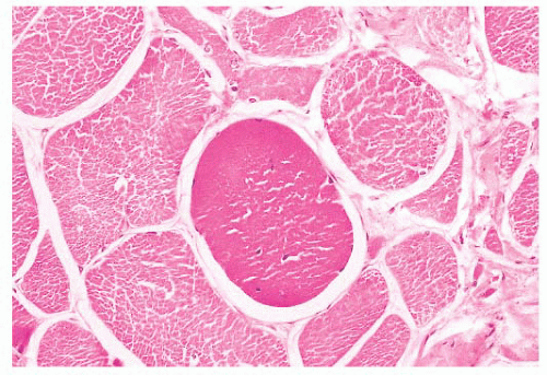
FIGURE 4.14 Hyaline fiber. The fiber in the center of the photograph is rounded, and it has dark, opaque sarcoplasm (H&E).


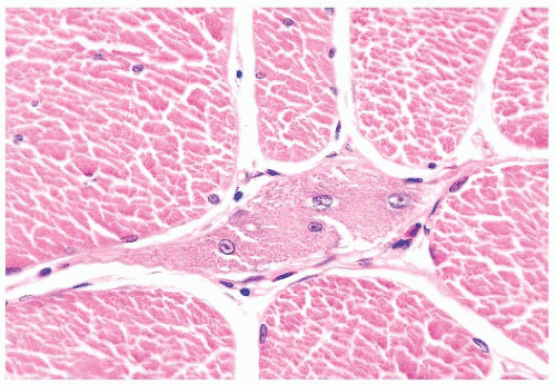
FIGURE 4.15 Regeneration. The nuclei of regenerating muscle fibers are large and vesicular with prominent nucleoli. The sarcoplasm is basophilic (H&E).


Fibrosis and Fatty Infiltration

Endomysial fibrosis is a common pathologic component of chronic necrotizing myopathies such as muscular dystrophies and myositis. The fibrosis is simply a manifestation of inflammation and repair as can be seen in most tissues. However, the factors that provoke end-stage interstitial fibrosis and fatty replacement of muscle have not been adequately explained by research investigations even though they are the bequest of chronic neuromuscular disease of both myopathic and neurogenic origin. Even the most seasoned pathologist experiences difficulty in interpreting the biopsy specimen with marked fibrosis or fatty infiltration because, at this juncture in the natural history of the disease, the active pathologic process has probably subsided and the opportunity of discovering specific pathologic changes is irretrievably lost (Fig. 4.16). Because end-stage muscle is unlikely to provide information relevant to the patient’s diagnosis, the biopsy of a severely involved muscle should be discouraged.


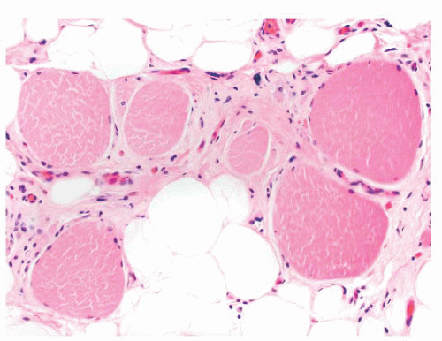
FIGURE 4.16 End-stage muscle. Atrophy, hypertrophy, pyknotic nuclear clusters, endomysial fibrosis, and fatty replacement are common features of end-stage muscle, regardless of the underlying neuromuscular disorder.


Ring Fibers

The ring is formed by a peripheral bundle of myofibrils that is directed circumferentially, encircling the inner portion of the myofiber, which otherwise appears normal in disposition and structure. In cross sections of muscle, the striated annulation is oriented in the transverse plane rather than in the longitudinal axis of the fiber (Fig. 4.17). The striations are easily visible in phosphotungstic acid hematoxylin (PTAH) or PAS-stained sections, resin sections, and ultrastructurally; with electron microscopy, the abnormally oriented myofibrils usually exhibit a normal architecture except for the hypercontraction of the sarcomeres (27). Although ring fibers have been reported in a variety of diseases, they are most consistently observed in limb-girdle dystrophy and myotonic dystrophy. Large numbers of ring fibers especially favor the latter diagnosis.


Inclusion Bodies

Inclusions may be located within the nuclei or within the sarcoplasm. Intranuclear inclusions suggest the diagnosis of oculopharyngeal dystrophy or inclusion body myositis. Sarcoplasmic inclusions suggest the diagnosis of myofibrillar myopathy or inclusion body myositis. In oculopharyngeal dystrophy, intranuclear inclusions are difficult to detect by light microscopy. Electron microscopic examination of the muscle tissue is likely to reveal nuclear inclusions composed of 8.5-nm unbranched tubular structures.

Nuclear inclusions in inclusion body myositis are faintly pink to wine colored in H&E stains, and they often fill the nucleus, leaving a rim of peripheral chromatin (Fig. 4.18). Pink or red inclusions may also be seen associated with the sarcoplasmic rimmed vacuoles (see Fig. 4.28). By electron microscopy, these inclusions, which consist of membranous whorls and bundles of filaments measuring 15 to 18 nm in diameter, may be present in few fibers, and they are extremely difficult to identify. The filaments are considered to be β-amyloid fibrils (see “Inclusion Body Myositis” section). Because the filaments are sparse, Congo red stains tend to be negative despite contrary claims in the literature.


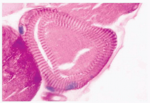
FIGURE 4.17 Ring fiber. Circumferential orientation of the peripheral myofibrils produces a striated ring that encircles a transversely sectioned muscle fiber (PAS).


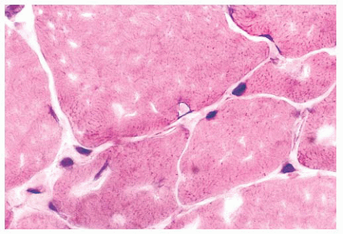
FIGURE 4.18 Intranuclear inclusion. An intranuclear inclusion is shown at the center of the photomicrograph. The inclusion is eosinophilic and smudged; it is located within a muscle fiber nucleus. The biopsy is from a patient with inclusion body myositis (H&E).

In the myofibrillar myopathies, some congenital myopathies, and miscellaneous other neuromuscular disorders, the inclusion bodies represent protein aggregates within the cytoplasm of affected muscle fibers. They may contain intermediate filaments such as desmin, which form poorly defined, smudged areas within the fibers. In hyaline body myopathy, the inclusions, which contain myosin filaments, are subsarcolemmal, well-defined, and slightly different in texture from the surrounding sarcoplasm. The protein composition of nonspecific cytoplasmic inclusion bodies has not been defined.

In contrast, nemaline rods are composed primarily of proteins normally found in Z-bands. Rods were first recognized in nemaline myopathy, a congenital and nonprogressive muscle disease of childhood (28). The name nemaline is derived from the Greek root nema (“thread” or “threadlike”) to emphasize the pathologic marker of this disorder. The threads or rods tend to cluster beneath the sarcolemma. Rods easily escape detection in H&E sections but are often readily apparent with Gomori trichrome stains of frozen sections (Fig. 4.19) or in toluidine blue-stained resin sections. Ultrastructurally, the rods are osmiophilic oblong or rectangular structures of varying dimensions, typically less than 5 µm (Fig. 4.20). Their latticelike appearance resembles that of the normal Z-band (29), likely a consequence of a protein composition that is similar to Z-bands. Since the original description of nemaline myopathy, it has become increasingly evident that rods are not unique to a single disease entity. Occasional rods in a small number of fibers have been reported in muscle specimens of muscular dystrophy and PM, for example. One cannot be comfortable with predicting the diagnosis of nemaline myopathy unless, in the appropriate clinical setting, many fibers in the specimen contain rods and they are numerous within each fiber.


Mottled Fibers and Lobulated Fibers

The peculiar, uneven staining reaction of mottled or moth-eaten fibers is satisfactorily demonstrated only in oxidative enzyme preparations. Zones of weak enzyme activity with irregular and poorly delimited borders are randomly dispersed in the sarcoplasm (Fig. 4.21). Ultrastructurally, mottled areas reveal a lack of mitochondria and the destruction of the myofilaments. The fact that the ultrastructural integrity of much of the cell sarcoplasm between the zones of mottling is preserved upholds the notion that the mottled fiber has the capacity for recovery and that it represents a form of reversible injury. Moth-eaten change is a nonspecific abnormality.


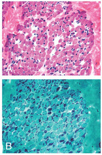
FIGURE 4.19 Nemaline myopathy. Nonspecific myopathic features of muscle fiber atrophy and hypertrophy are typically the predominant pathology in H&E stained sections (A). Collections of dark, granular to rod-shaped inclusions are striking after staining with Gomori trichrome (B).

Lobulated fibers have a superficial resemblance to mottled fibers (see preceding paragraph) and fibers containing minicores (see next paragraph). Unlike moth-eaten changes and cores, lobulations are easily recognized on H&E and Gomori trichrome (Fig. 4.22) as well as oxidative stains. The peripheral cytoplasm of muscle fibers is divided by thin triangular clusters of mitochondria that extend a short distance centrally from the sarcolemma. Although generally considered to be a feature of myopathic disease, lobulated fibers do not have disease specificity.


Cores and Targets

Oxidative enzyme reactions are the ideal technique for identifying cores that appear as sharply demarcated regions of
depleted or absent mitochondria (Fig. 4.23). Large cores are often also evident with H&E and Gomori trichrome stains. Ultrastructurally and in resin sections, distinguishing between structured and unstructured cores is possible (30). The crossbanding pattern is evident in the structured core, whereas cross-striations are absent from the unstructured core, which perhaps represents a later stage in core development. Cores cannot be regarded as a specific pathologic finding because they occur in a variety of diseases. However, the muscle fibers of several inherited myopathies contain cores, and one nosologic group, the core myopathies, is defined by them. As a nonspecific change, cores are present in less than 10% of fibers, whereas they are numerous and are located more often in type 1 fibers in central core disease. Although cores are single and centrally placed within the fiber in classic central core disease, they may be multiple and eccentric in other conditions.


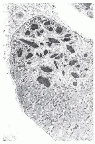
FIGURE 4.20 Nemaline myopathy ultrastructure. Nemaline rods are osmiophilic, resembling the electron density of Z-bands (electron micrograph).


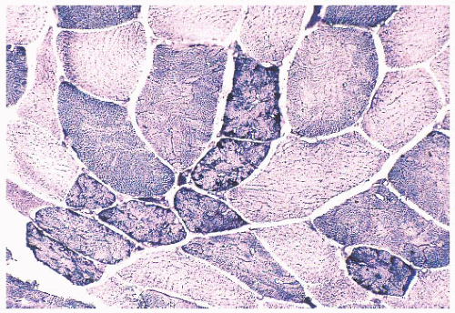
FIGURE 4.21 Mottled fibers. The sarcoplasm appears moth eaten as a result of the presence of patchy areas of poor staining (NADH).


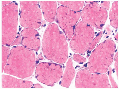
FIGURE 4.22 Lobulated fibers. Several muscle fibers in this image are divided into lobules by septa occupied by mitochondria (H&E).

For diagnostic purposes, target fibers are considered pathognomonic for neurogenic atrophy (31), but unfortunately, they are identified in less than 25% of cases. Despite many similarities, targets and cores can often be distinguished. Of greatest significance is the three-zone structure of the target. The central zone, which resembles the unstructured core, is surrounded by an intermediate zone, which is darkly stained in oxidative enzyme reactions (Fig. 4.24). This rim, which is not a feature of cores, sharply contrasts with the third zone, the
outer normal portion of the fiber. The term targetoid refers to target-like, sharply demarcated regions that lack the intermediate zone or rim of increased oxidative enzyme activity. Targetoid change and cores essentially are morphologically identical, but the term targetoid is typically used in neuropathic disease and cores in myopathic disease.


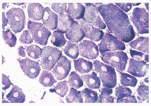
FIGURE 4.23 Cores. Muscle fibers contain cores (sharply demarcated areas of reduced enzyme activity) in several types of inherited myopathies. In this patient with central core myopathy, single cores are present within many muscle fibers (NADH).


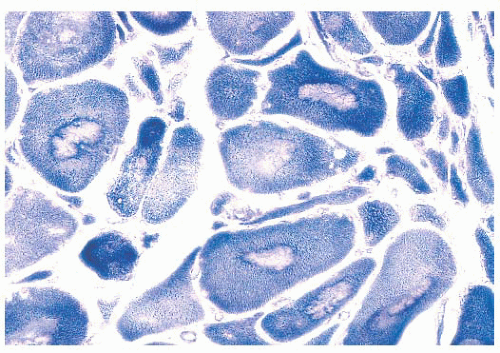
FIGURE 4.24 Target fibers. In target fibers, an inner, unstained zone is surrounded by a rim of increased enzyme activity (NADH). Targets are seen in the context of neurogenic atrophy, and angulated atrophic fibers are present in this image.


Mitochondrial Abnormalities

Abnormalities of mitochondria occur in a wide variety of diseases (32,33 and 34). They may be generalized disorders, or they may be limited to skeletal muscle. The mitochondrial abnormalities are similar in all of these diseases, and they are often recognized by the presence of ragged red fibers. Classic ragged red fibers are readily identified by the Gomori trichrome, in which intensely red, subsarcolemmal protrusions from the cell surface give the margins of involved fibers an irregular, ragged appearance (Fig. 4.25). Such fibers are surrounded by prominent, sometimes dilated capillaries that appear to indent them and to be increased in number. Ragged red fibers are equally impressive in oxidative enzyme reactions, especially succinate dehydrogenase reactions, in which large collections of mitochondria are seen as dark, coarsely granular deposits that not only may be subsarcolemmal but may also diffuse throughout the fiber.


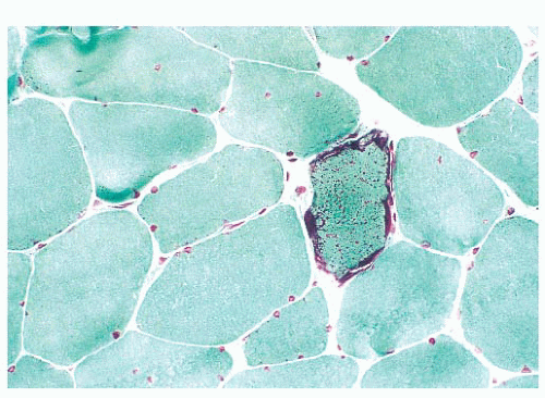
FIGURE 4.25 Ragged red fiber. Collections of mitochondria appear as red-stained, irregular, subsarcolemmal areas within the involved fiber (Gomori trichrome).

Electron microscopy shows that ragged red fibers contain accumulations of mitochondria that may be enlarged and abnormal in shape. Their cristae may be excessively numerous and disorganized, or they may be concentrically arranged. Among the commonly encountered matrix inclusions are glycogen aggregates, clear vacuoles, floccular densities, myelin figures, and paracrystalline structures having a square or rectangular conformation and resembling a parking lot or grid (Fig. 4.26). These inclusions contain mitochondrial creatine kinase and perhaps other proteins.


Vacuolar Change

Vacuoles may contain abnormal quantities of glycogen or lipid, or they may be composed of clustered lysosomes/autophagosomes (Table 4.7). Hence, routine microscopic sections with vacuolar change of an unexplained nature should be stained with PAS and oil red O or other suitable fat stains. We prefer to use resin sections, in which the osmiophilic fat deposits are more clearly demonstrated than they are in routine fat stains (Fig. 4.27). An abundance of lipid within myofibers is a relatively specific finding that is usually indicative of a lipid storage disease or mitochondrial myopathy, with the latter frequently being recognized by the presence of ragged red fibers. Although deposits of glycogen raise the specter of carbohydrate storage disease or other disorders affecting glycogen metabolism, such as hypothyroidism (35), they can be an incidental, fortuitous finding in virtually any disorder. Especially when only one or two fibers in the specimen are involved and the vacuoles are crescentlike, subsarcolemmal,
and visible primarily in PAS stains, they are probably of no clinical significance.


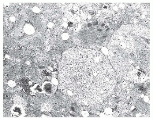
FIGURE 4.26 Mitochondrial myopathy. Ultrastructurally, a ragged red fiber shows numerous mitochondria. Here, some of the mitochondria are abnormally large, and some contain paracrystalline inclusions (electron micrograph).


TABLE 4.7 Sarcoplasmic Vacuoles Seen in the Biopsy Specimen


Pathologic Features


Disease


Often center of specimen, often arranged in size gradient


Freezing (ice crystal) artifact


Often subsarcolemmala


Glycogen storage (McArdle disease)


Multiple, throughout the muscle fibera


Glycogen storage (Pompe disease)


In scattered fibers; small, round, osmiophilic; oil red O positive


Lipid storage or mitochondrial myopathies


Rimmed by granular, ubiquitin-positive material


IBM, distal myopathies, OPMD, hydroxychloroquine toxicity


Rimmed by membrane expressing sarcolemmal proteins


Autophagic vacuolar myopathies (e.g., Danon disease and XMEA)


a The vacuoles in glycogen storage diseases are typically empty in routine cryosections and paraffin sections because the glycogen is lost during processing. Sometimes, there is residual PAS-positive material. More reliably, there is slightly metachromatic material filling these vacuoles in epon (plastic) sections stained with toluidine blue.


IBM, inclusion body myositis; OPMD, oculopharyngeal muscular dystrophy; XMEA, X-linked myopathy with excess autophagy.


The so-called rimmed vacuole has received attention as a diagnostic criterion of oculopharyngeal dystrophy (36), distal myopathy (37), and inclusion body myositis (38). These vacuoles may be sharply demarcated and may contain granular material (Fig. 4.28). Granular material typically rims the vacuoles, appearing basophilic in H&E stains and red in Gomori trichrome stains of frozen sections. Ultrastructurally, this type of vacuole is not always enclosed by a membrane, and it contains lysosomal membranous profiles with or without tubulofilamentous material. The rimmed vacuole is believed to be derived
from the autophagic vacuole, which classically is membrane bound and is a repository for the debris of autodigestion. In at least two genetic diseases, autophagic vacuoles are numerous (39,40). These vacuoles are rimmed by membrane that expresses proteins typically found in the sarcolemma. Enzyme histochemistry or immunostaining for these sarcolemmal proteins demonstrate strikingly numerous cytoplasmic vacuoles in Danon disease and X-linked myopathy with excessive autophagy (XMEA) (see Fig. 4.36).


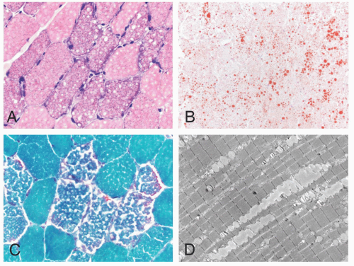
FIGURE 4.27 Lipid storage myopathy. Numerous vacuoles are evident in the sarcoplasm of affected muscle fibers (A, H&E; C, Gomori trichrome). The vacuoles contain neutral lipid that can be demonstrated by histochemistry (B, oil red O) or by electron microscopy (D).


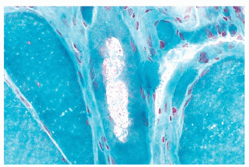
FIGURE 4.28 Rimmed vacuole. Aggregates of lysosomes have the appearance of rimmed vacuoles in frozen sections. The muscle fiber in the center contains rimmed vacuoles with red, granular material (Gomori trichrome).


Changes in Fiber Type Distribution

The normal checkerboard staining paradigm that is so familiar in histoenzymatic reactions may fall victim to certain pathologic conditions. Predominance of one fiber population is emblematic of the congenital myopathies. Type 1 fiber predominance is found in central core disease, nemaline myopathy, centronuclear myopathy, and some cases of congenital fiber-type disproportion. In chronic denervation, the normal checkerboard pattern is replaced by large groups of fibers with identical histochemical properties. This fiber type grouping (Fig. 4.11) is a result of reinnervation of the denervated muscle. Normal anatomic and physiologic principles help to explain type grouping. The muscle fibers in each motor unit are of a single histochemical type. The neuron, through its axon and intramuscular branches (nerve twigs), which innervate the individual fibers, governs the fiber type in the motor unit. Reinnervation of a type 1 fiber by a type 2 nerve twig has been shown experimentally to convert the fiber from type 1 to type 2. The opposite occurs with a type 2 fiber and a type 1 twig. Thus, when neighboring denervated fibers of differing histochemical types undergo reinnervation by collateral sprouts from a single axon that has remained viable, all of the fibers are converted to one histochemical type, and fiber type grouping results.


NEUROMUSCULAR DISEASES

The muscle biopsy is only one facet of the overall diagnostic evaluation of the patient. It must not be interpreted in a vacuum without a consideration of the clinical history, physical examination findings, and the results of all pertinent laboratory tests. This section provides the background needed to interpret the biopsy findings in the context of the clinical setting. Those neuromuscular diseases that are most often encountered and in which the muscle biopsy significantly contributes to the diagnosis are emphasized. Based on more than 25 years of experience at the University of Buffalo and the University of Iowa, the most common neuromuscular diseases evaluated by muscle biopsy are either inflammatory or neurogenic. Despite best efforts, a specific diagnosis cannot be reached in some biopsies, so the pathology reports must be descriptive without a definitive interpretation.


MUSCULAR DYSTROPHIES

The rather meaningless term dystrophy, which literally means “deficient nutrition,” was popularized toward the close of the nineteenth century, when the pathogenesis of the muscular dystrophies was totally mysterious. The muscular dystrophies, which share certain clinical and pathologic attributes, have long been considered part of a common rubric. In general, the initial symptoms are manifested during childhood or young adulthood; however, late adult onset cases do occur. The cardinal symptom is muscular weakness that is steadily and unremittingly progressive. By definition, all forms of muscular dystrophy are genetic (Table 4.8); some are inherited, whereas others are de novo mutations. The histopathologic criteria of muscle fiber necrosis (myonecrosis), regeneration, and endomysial fibrosis are common to all patients (Fig. 4.29), but the severity of dystrophic changes varies among genotypes, among allelic variants of the same genotype, among muscles within individual patients, within a single biopsy, and over time within individual patients. Specific genetic diagnoses can be suggested by immunostaining, but definitive diagnoses rely on molecular genetic testing.


Dystrophinopathy (Duchenne and Becker Muscular Dystrophies)

Duchenne muscular dystrophy (DMD) is the most common form of muscular dystrophy and also among the most severe. The dystrophin gene (DMD) that is responsible for DMD is located on the short arm of the X chromosome at Xp21 (41). With a 14-kb coding sequence within 2.5 to 3.0 Mb of DNA, it is the largest known human gene. Approximately two-thirds of DMD cases are caused by large deletions or duplications that are easily detected by genomic hybridization (42). Complete sequencing of coding exons and exon/intron boundaries discovers all but about 2% of the remaining DMD mutations (43). The identification of mutations permits an accurate diagnosis, including carrier and prenatal detection. Patients with DMD are greatly deficient in full-length dystrophin, a 427-kd, rod-shaped protein that is found predominantly in skeletal and cardiac muscle (44,45). Dystrophin is localized just beneath the sarcolemmal membrane and is a key component of the dystrophin-glycoprotein complex (DGC; Fig. 4.30). The DGC is presumed to promote sarcolemmal stability, particularly during muscle contraction, by forming a structural alignment between cytoskeletal actin inside and extracellular matrix proteins (e.g., merosin [laminin α2 or laminin 211], perlecan, and agrin) within the basement membrane (46). Other key members of the DGC are dystroglycans and sarcoglycans (see Fig. 4.30 and sections on limb-girdle and congenital muscular dystrophies).


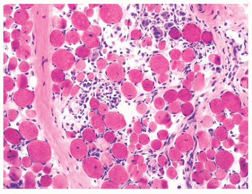
FIGURE 4.29 Muscular dystrophy histopathology. The classic histopathologic features of muscular dystrophies are illustrated here: myonecrosis and regeneration without significant lymphocytic inflammation, internally placed nuclei, muscle fiber atrophy or hypertrophy, and endomysial fibrosis (H&E).


TABLE 4.8 Inherited Myopathiesa


Disease


Gene (Protein)


Chromosome


X-LINKED MUSCULAR DYSTROPHIES


Dystrophinopathies (Duchenne, Becker, and manifesting carriers)


DMD (dystrophin)


Xp21


Emery-Dreifuss muscular dystrophy (EDMD)


EMD (emerin)


Xq28


AUTOSOMAL DOMINANT MUSCULAR DYSTROPHIES


Facioscapulohumeral dystrophy (FSHD)


Complex genetic mechanism involving DUX4 (double homeobox 4)


Deletion of D4Z4 repeats at 4q35


Myotonic dystrophy, type 1 (DM1)


DMPK (myotonic dystrophy protein kinase)


19q13


Myotonic dystrophy, type 2 (DM2)


CNBP (CCHC-type zinc finger nucleic acid-binding protein; formerly ZNF9, zinc finger protein 9)


3q21


Oculopharyngeal muscular dystrophy


PABP2 (poly A binding protein, nuclear 1)


14q11


Limb-girdle muscular dystrophies (LGMD 1)


LGMD 1A


MYOT (myotilin)


5q31


LGMD 1B (also dominant EDMD)


LMNA (laminA/C)


1q21


LGMD 1C


CAV3 (caveolin3)


3p25


LGMD 1D


DNAJB6 (HSP-40 homologue, subfamily B, number 6)


7q36


LGMD 1E


DES (desmin)


2q35


LGMD 1F


TNPO3 (transportin 3)


7q32


AUTOSOMAL RECESSIVE MUSCULAR DYSTROPHIES


Congenital muscular dystrophies (CMD)


Merosin-deficient CMD (MDC1A)


LAMA2 (laminin α2)


6q22


Dystroglycanopathies – genetically and phenotypically heterogeneous muscular dystrophies; at least 15 genes published by summer 2013 involved in the O-mannosylation of α-dystroglycan; most severe congenital forms involve muscle, eye, and brain (e.g., Walker-Warburg syndrome, muscle-eye-brain disease, and Fukuyama congenital muscular dystrophy); other congenital forms include MDC1C (FKRP mutations) and MDC1D (LARGE mutations); milder allelic forms include LGMD subtypes 2I, 2K, 2M, 2N, and 2O (see the following)


Collagen VI-related dystrophies (Ullrich CMD)


COL6A1, A2 or A3 (alpha1, alpha2, or alpha3 chains of type VI collagen)


21q22 2q37


Megaconial CMD


CHKB (choline kinase beta)


22q13


Limb-girdle muscular dystrophies (LGMD 2)


LGMD 2A


CAPN3 (calpain-3)


15q15


LGMD 2B


DYSF (dysferlin)


2p12


LGMD 2C


SCGC (γ-sarcoglycan)


13q12


LGMD 2D


SCGA (α-sarcoglycan)


17q21


LGMD 2E


SCGB (β-sarcoglycan)


4q12


LGMD 2F


SCGD (δ-sarcoglycan)


5q33


LGMD 2G


TCAP (telethonin)


17q12


LGMD 2H


TRIM32 (tripartite motif-containing 32)


9q33


LGMD 2I


FKRP (fukutin-related protein)


19q13.3


LGMD 2J


TTN (titin)


2q31


LGMD 2K


POMT1 (protein-O-mannosyltransferase 1)


9q34


LGMD 2L


ANO5 (anoctamin 5)


11p14


LGMD 2M


FKTN (fukutin)


LGMD 2N


POMT2 (protein-O-mannosyltransferase 2)


14q24


LGMD 2O


POMGnT1 (O-linked mannose beta1,2-N-acetylglucosaminyltransferase)


1p34


LGMD 2Q


PLEC1 (plectin 1)


8q24


CONGENITAL MYOPATHIES


Core diseases (dominant or recessive)


RYR1 (ryanodine receptor 1)


19q13


SEPN1 (selenoprotein N1)


1p36


Nemaline myopathies (dominant or recessive)


sarcomeric thin filaments (ACTA1, CFL2, KBTBD13, NEB, TNNT1, TPM2, TPM3)


multiple loci


Congenital fiber-type disproportion (CFTD; dominant or recessive)


(ACTA1, MYH7, RYR1, SEPN1, TPM3)


multiple loci


Centronuclear myopathies (X-linked, dominant, or recessive)


MTM1 (myotubularin)


BIN1 (amphiphysin; recessive)


DNM2 (dynamin 2; dominant)


Xq28


2q14


19p13


OTHER INHERITED MYOPATHIES


Danon disease (X-linked)


LAMP2 (lysosome-associated membrane protein 2)


Xq24


X-linked myopathy with excess autophagy (XMEA)


VMA21 (vacuolar ATPase assembly integral membrane protein VMA21)


Xq28


Distal myopathies (dominant; inclusion body myopathy with early onset Paget disease and frontotemporal dementia, IBMPFD; Welander distal myopathy)


VCP (valosin-containing protein)


TIA1 (RNA-binding protein “T-cell restricted intracellular antigen 1”)


9p13


2p13


Distal myopathies (recessive; Miyoshi myopathy; Nonaka myopathy)


Miyoshi – ANO5 or DYSF


Nonaka – GNE


11p14 or 2p12


9p13


Myofibrillar myopathies (dominant)


CRYAB, DES, MYOT, FLNC, BAG3, LDB3


Multiple loci


a Online resources include GeneTable (http://www.musclegenetable.fr/), GeneReviews (http://www.ncbi.nlm.nih.gov/books/NBK1116/), and Online Mendelian Inheritance in Man (OMIM; http://www.ncbi.nlm.nih.gov/omim)


IBMPFD, inclusion body myopathy with early-onset Paget disease of bone and frontotemporal dementia.

Only gold members can continue reading. Log In or Register to continue

Stay updated, free articles. Join our Telegram channel

Sep 22, 2016 | Posted by in PATHOLOGY & LABORATORY MEDICINE | Comments Off on Muscle Biopsy in Neuromuscular Diseases

Full access? Get Clinical Tree

Get Clinical Tree app for offline access