Neuroendocrine and Paracrine Systems


Neuroendocrine and Paracrine Systems


Ricardo V. Lloyd

Lori A. Erickson


This chapter is divided into two parts. The first part outlines the neuroendocrine system and presents an overview of the diffuse or dispersed neuroendocrine system (DNS) to help the general surgical pathologist gain a panoramic view of this complex system. Methods of analysis of tumors of the DNS are also presented because some of these techniques are indispensable for the diagnosis of many neuroendocrine neoplasms. The second part of the chapter discusses examples of specific lesions of the DNS. Some of the less common DNS tumors are presented, along with differential diagnoses, because the more common lesions are covered in detail in other areas of the text.

The DNS consists of a wide variety of cells that are present in the central and peripheral nervous system and in many classic endocrine organs (Table 11.1). The cells of the DNS have also been referred to historically as paraneurons (1). These cells share the ability to produce many biologically active amines, peptides, and other substances. These substances may act as neurotransmitters, as true hormones, or as paracrine regulators. Paracrine regulation refers to the production of amines and hormones by cells that exert a local effect on the target cells by diffusion through the extracellular space. The production of somatostatin by the pancreatic islets, which regulates insulin and glucagon production in neighboring islet cells, is an example of paracrine regulation. The cells and neoplasms of the neuroendocrine and paracrine systems make up the DNS.


DEVELOPMENT OF THE DISPERSED NEUROENDOCRINE SYSTEM CONCEPT

Feyrter (2) considered the clear cells of the gastrointestinal tract to be peripheral endocrine or paracrine cells. The detailed study of neuroendocrine cells by Pearse (3,4) led to the development of the concept of amine precursor uptake and decarboxylation (APUD). Although the APUD concept provided a unifying theory for explaining some endocrine diseases and ectopic hormone productions, the hypothesis that the cells were all of neural crest origin, as postulated by Pearse, was later disproved by the experiments of LeDouarin (5) and others. The current neuroendocrine classification of cells and tumors uses immunohistochemical (IHC), ultrastructural, and molecular biologic features to define members of the DNS (6,7 and 8).


CELLS AND NEOPLASMS OF THE DISPERSED NEUROENDOCRINE SYSTEM

The principal cells and neoplasms that form the DNS are listed in Table 11.1. The steroid-producing endocrine cells of the adrenal cortex, ovary, and testis, as well as the thyroid hormone-producing follicular cells in the thyroid gland, do not form part of the DNS. Cells and neoplasms of the DNS may be divided into the following two principal groups: (a) those of neural type, which include neuroblastomas, pheochromocytomas, and paragangliomas; and (b) those of epithelial type, which include neuroendocrine neoplasms from many sites. Many of these neoplasms have distinct clinicopathologic features, so precise classification by the pathologist is necessary for optimal clinical management. Although the term carcinoid tumor has been broadly used to refer to many neoplasms derived from the DNS (9), this term is being replaced by neuroendocrine neoplasms. However, to communicate effectively with clinicians, the term carcinoid tumor can still be used in parenthesis for neoplasms of the gastrointestinal tract and lungs. The most recent World Health Organization 2010 recommendation for the general endocrine tumor classification includes (10):


  • Neuroendocrine tumor (NET), G1 (carcinoid) or G2


  • Neuroendocrine carcinoma (NEC), G3, large cell or small cell type


  • Mixed adenoneuroendocrine carcinoma (MANEC)


  • Hyperplastic and preneoplastic lesions

Many broad-spectrum IHC markers are available to aid in the diagnosis of neuroendocrine neoplasms. The principal markers include chromogranins and synaptophysin. Other markers that can be helpful but are of more limited use include neuron-specific enolase, proconvertases PC1/PC3 and PC2, bombesin and/or gastrin-releasing peptide (GRP), CD57 (Leu-7/HNK-1), synaptic vesicle protein 2, PGP9.5, and others (11). Although historical silver stains, such as Grimelius (12) and Churukian-Schenk (13) argyrophilic stains (a histochemical reaction in which the endocrine cells take up silver ions, but a reducing agent is needed to produce a positive reaction) and the Masson-Fontana argentaffin stain (a histochemical reaction in which the endocrine cells take up and reduce silver ions without a reducing agent), are helpful in characterizing some neuroendocrine neoplasms, the great variability in silver stains leads to less consistent results than those yielded by IHC stains. However, IHC stains are not without problems.
Interlaboratory variability resulting from use of different antibodies and different techniques remains a serious problem. Despite this variability, the predictability and reproducibility of standardized IHC reagents, such as monoclonal antibodies, has made the use of these reagents to characterize neuroendocrine neoplasms widely accepted.


TABLE 11.1 Cells and Neoplasms of the Dispersed Neuroendocrine Systema


Cell/Tissue


Neoplasms


Hormones/Amines


Adrenal medulla


Pheochromocytoma, neuroblastoma, and ganglioneuroma


Enkephalins, catecholamines, VIP, and SRIF


Biliary tract and liver


Benign and malignant tumors


Serotonin, catecholamines, and miscellaneous peptides


Bronchopulmonary tree


Neuroendocrine tumors (including “carcinoids”)


GRP, ACTH, endorphin, calcitonin, catecholamines, and serotonin


Gastrointestinal tract


Benign and malignant tumors (including “carcinoids”)


Gastrin, SRIF, GRP, VIP, secretin, enteroglucagon, PP, serotonin, catecholamines, cholecystokinin, insulin, and glucagon


Hypophysis


Adenomas and carcinomas


ACTH, GH, FSH, LH, PRL, TSH, β-endorphin, calcitonin, catecholamines, and MSH


Merkel cells of skin


Neuroendocrine (Merkel cell) carcinoma


Calcitonin, catecholamines, PP, and VIP


Pancreatic islet


Benign and malignant tumors


Insulin, PP, glucagon, SRIF, VIP, gastrin, catecholamines, and serotonin


Paraganglion


Paragangliomas


Enkephalin, catecholamines, and VIP


Parathyroid


Adenomas and carcinomas


Parathyroid hormone catecholamines


Thyroid C cell


Medullary thyroid carcinoma


Calcitonin, somatostatin, ACTH, CGRP, and catecholamines


Miscellaneous neuroendocrine cells in breast, cervix, kidney, larynx, ovary, paranasal sinus, prostate, testis, and other sites


Benign and malignant tumors and mixed tumors


Miscellaneous peptides, serotonin, and catecholamines


aThe hypothalamus and pineal gland are also members of the dispersed neuroendocrine system.


ACTH, adrenocorticotropic hormone; CGRP, calcitonin gene-related peptide; FSH, follicle-stimulating hormone; GH, growth hormone; GRP, gastrin-releasing peptide; LH, luteinizing hormone; MSH, melanocyte-stimulating hormone; PP, pancreatic polypeptide; PRL, prolactin; SRIF, somatostatin; TSH, thyroid-stimulating hormone; VIP, vasoactive intestinal polypeptide.


GENERAL NEUROENDOCRINE MARKERS


CHROMOGRANIN/SECRETOGRANIN

The chromogranin/secretogranin (Cg/Sg) family is composed of several acidic proteins that are present in the secretory granules of neuroendocrine cells. The three major Cg/Sg proteins are currently designated as chromogranin A, B, and secretogranin II (Sg II). The distribution of chromogranin A has been studied extensively in human tumors (14). It is present in most neuroendocrine cells and neoplasms. However, most neoplasms with only a few endocrine secretory granules, such as small cell carcinomas of the lung, do not react strongly with chromogranin A antibodies (14). Because of their widespread distribution and high degree of specificity, chromogranins A and B and Sg II are excellent markers for neuroendocrine cells and neoplasms (15,16).


SYNAPTOPHYSIN

This 38-kd molecule is a component of the membrane of presynaptic vesicles. It is widely distributed in neurons and neuroendocrine cells and their neoplasms, and it is another broad-spectrum neuroendocrine marker that is localized in cytoplasmic vesicles rather than in secretory granules (15,17). Some endocrine tumors that are not members of DNS, such as adrenal cortical adenomas and carcinomas, may express synaptophysin; thus, this marker should be used with other neuroendocrine markers, such as chromogranin. Many other related vesicle proteins, such as synaptobrevin, syntaxin, synaptogranin, SNAP-25B, and rabphilin 3A, have been found to be associated with cytoplasmic vesicles (18). However, their clinical use and specificity as broad-spectrum neuroendocrine markers have not been demonstrated.


NEURON-SPECIFIC ENOLASE

The neuron-specific enolase (NSE) enzyme, which is also known as γ-enolase, is a highly sensitive (but not too specific) marker for neuroendocrine cells and tumors. It is commonly found in neurons, peripheral nerves, and neuroendocrine cells (19). Some nonneuroendocrine cells and neoplasm also react with antisera against NSE. In the diagnosis of NETs, NSE should be used only with other broad-spectrum markers of neuroendocrine cells.


PROCONVERTASES

The proconvertases (PCs) are recently described enzymes that process propeptides into active peptides within cells. Some of these, including PC1/PC3 and PC2, are highly specific for neuroendocrine cells and tumors (20), and they can be used as specific neuroendocrine markers.


TABLE 11.2 Sites of Eutopic and Ectopic Hormone Production of Common Neuroendocrine Hormones


Hormone


Produced by Cells/Tumors


Other Sites of Production


Calcitonin


C cell/medullary thyroid carcinoma


Laryngeal NE tumors; pancreatic NE tumors


ACTH


Pituitary/pituitary adenomas


Pancreatic NE tumors; lung NE tumors; pheochromocytomas


CRH


Hypothalamic neuromas


Pancreatic NE tumors


GH


Pituitary/pituitary adenomas


Pancreatic NE tumors


GHRH


Hypothalamic neuromas


Pancreatic NE tumors


Pancreatic polypeptide


Pancreatic islet/pancreatic NE tumors


Intestinal NE tumors (hindgut carcinoids)


Somatostatin


Pancreatic islet/pancreatic NE tumors, duodenum, stomach


Intestinal NE tumors (hindgut carcinoids)


ACTH, adrenocorticotropic hormone; CRH, corticotropin-releasing hormones; GH, growth hormone; GHRH, growth hormone-releasing hormone; NE, neuroendocrine.


BOMBESIN/GASTRIN-RELEASING PEPTIDE AND CD57 (LEU-7/HNK-1)

Bombesin is a tetradecapeptide originally isolated from amphibian skin. It is present in many endocrine cells as well as in central and peripheral neurons (21). GRP, the proposed mammalian analogue of bombesin, has been found in many lung and gastrointestinal endocrine tumors (22) and can be used as a broad-spectrum marker for many endocrine neoplasms.

CD57 (Leu-7/HNK-1), a monoclonal antibody that was produced against a T-cell leukemia cell line, recognizes natural killer cells in blood and lymphoid tissues. It also reacts with small cell carcinomas of the lung as well as with pheochromocytomas and other neuroendocrine neoplasms (23,24).


OTHER IMMUNOHISTOCHEMICAL MARKERS

With advances in proteomics, newer neuroendocrine markers are being discovered. A promising new marker is synaptic vesicle protein 2, which is associated with secretory granules and which should serve as a good complement to chromogranin and synaptophysin (25). Many other IHC markers, such as PGP9.5, antibodies against enzymes in the synthetic pathways for peptide hormones, and CD56 (neural cell adhesion molecules), have been used to characterize some neuroendocrine cells and tumors (13). However, these are generally used as second or adjunctive markers in problematic cases.


REGULATORY PEPTIDES AND AMINES

A broad spectrum of peptides and amines is present in cells and tumors of the DNS (see Table 11.1). Although the biologic function of many of these is known, some of these substances are without known activity. The localization of peptides may be of some help in characterizing certain neuroendocrine neoplasms. However, ectopic production of peptides is a common phenomenon, so the presence of a specific peptide may not be helpful in characterizing unusual neuroendocrine neoplasms. Although most NETs express broad-spectrum markers such as chromogranin and synaptophysin, specific peptides are not commonly found in specific tumor types. For example, calcitonin is frequently expressed by medullary thyroid carcinomas and atypical laryngeal carcinoids but may also be expressed ectopically in other tumors (Table 11.2).


ULTRASTRUCTURAL CHARACTERISTICS OF NEUROENDOCRINE CELLS AND NEOPLASMS

When the differential diagnosis of an anaplastic neoplasm includes a neuroendocrine carcinoma, the presence of secretory granules on ultrastructural examination can be helpful in establishing the diagnosis of a malignant neuroendocrine neoplasm. Cytoplasmic secretory granules usually have a central or eccentric core of variable density and limiting membrane. The size of the secretory granules ranges from 50 to 400 nm (Fig. 11.1). Although the morphology of secretory granules in many normal neuroendocrine cells can be used as an aid in recognizing specific cell types, these distinct morphologic granule features are not often seen in neuroendocrine neoplasms. Recent studies have shown that several hormones and amines, such as calcitonin and somatostatin, may be stored in the same secretory granules in medullary thyroid carcinomas (26) and that Cg/Sg proteins form a major constituent of the secretory granule (14,15).


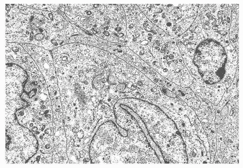
FIGURE 11.1 Neuroendocrine carcinoma of lung from a patient with Cushing syndrome. The carcinoma was positive for adrenocorticotropic hormone and corticotropin-releasing hormone by immunostaining. Neurosecretory granules ranging in size from 100 to 300 nm in diameter are present in the cytoplasm (8600×).


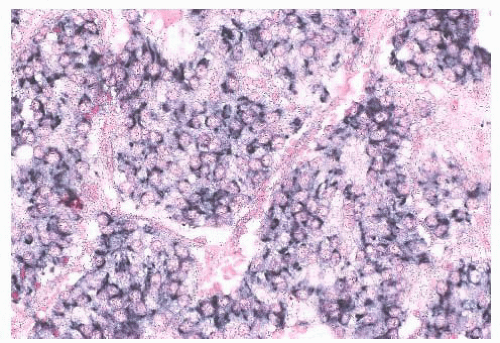
FIGURE 11.2 In situ hybridization localizing chromogranin A RNA diffusely in a retroperitoneal neuroendocrine tumor (in situ hybridization with a digoxigenin-labeled probe cocktail of chromogranin A and B with streptavidin nitroblue tetrazolium-5-bromo-4-chloro-3-indolyl phosphate [NBT-BCIP] detection).


IN SITU HYBRIDIZATION AND POLYMERASE CHAIN REACTION

Localization of the mRNA for specific peptides and other neuroendocrine markers by in situ hybridization (ISH) is another useful technique for characterizing neuroendocrine cells and neoplasms (8,27). Some neuroendocrine neoplasms may contain mainly the mRNA but not the translated product for specific hormones, so detection of the mRNA within the cells can help to establish the diagnosis (27) (Fig. 11.2). However, most NETs contain enough peptides, hormones, or broad-spectrum markers to be detected by IHC techniques. A notable exception is the production of ectopic hormones by NETs, in which ISH offers some advantages because of the retention of intracytoplasmic mRNA. The use of the polymerase chain reaction (PCR) to amplify small amounts of DNA and of reverse transcriptase-PCR to amplify specific products starting with RNA is becoming more common in diagnostic pathology (28,29). A combination of PCR and ISH has helped to amplify and to visualize specific RNA and DNA targets that are expressed in low abundance in tissue sections (30).


MULTIPLE ENDOCRINE NEOPLASIA SYNDROMES

The development of hyperplasia and neoplasms involving multiple organs of the DNS is usually a familial condition. The term multiple endocrine neoplasia (MEN) is applied to these syndromes. Several distinct patterns of familial MEN are observed; these are inherited as autosomal dominant traits with a high degree of penetrance. In type 1, the principal organs affected include the pituitary, pancreas, and parathyroid (31,32). Some patients may also have the Zollinger-Ellison syndrome with peptic ulceration. Type 2A usually involves the thyroid C cells and the adrenal medulla as well as the parathyroids (33). Type 2B also involves the thyroid C cells and adrenal medulla, and it is associated with mucosal neuromas (33). A new variant of the MEN syndrome includes mutations in the p27 (Kip1) and p18 (INK4C) genes and has been designated as MEN4 (34). Abnormalities of the parathyroid glands and pituitary glands are also involved with this syndrome. The studies of DeLellis and Wolfe (26) have shown that hyperplasias usually precede neoplasias in MEN syndromes involving the C cells of the thyroid and the adrenal medulla. The genes for MEN1 (MENIN) and MEN2A and 2B (rearranged during transfection [RET] proto-oncogene) have been characterized extensively (32). The availability of these genetic markers has facilitated screening and early diagnosis of MEN2A and 2B in affected families (35), although no general screening tests for MEN1 or MEN4 are available to date (34,36).


ECTOPIC HORMONE PRODUCTION

Ectopic or inappropriate hormone secretion is characterized by the production of hormones by a tumor in which the parent tissue from which the tumor was derived does not produce the hormone. The DNS provides a general unifying concept explaining ectopic hormone production by many endocrine tumors. Some non-NETs, such as squamous cell carcinomas of the lungs, hepatocellular carcinomas, and some sarcomas, may be associated with ectopic hormone production (37). Ectopic hormone production by some neoplasms, such as the production of adrenocorticotropic hormone (ACTH) by pancreatic endocrine neoplasms, may be associated with a more biologically aggressive tumor (38) (see Table 11.2). One of the most commonly produced ectopic hormones associated with hypercalcemia is parathyroid hormone-related protein (39). This protein is produced by parathyroid neoplasms as well as by many neuroendocrine and other tumors, and it is a major cause of hypercalcemia associated with malignancy (39).

Many of the common neuroendocrine neoplasms are discussed elsewhere in this book, so they are mentioned in this chapter only in discussions or differential diagnoses. These include pheochromocytomas, paragangliomas, medullary thyroid carcinomas, melanomas, pancreatic and gastrointestinal neuroendocrine neoplasms, and neuroendocrine neoplasms of the lungs. The terminology used in this chapter is from the most recent World Health Organization recommendation for general endocrine tumor classification (10).


NEUROENDOCRINE NEOPLASMS


THYMUS

Thymic neuroendocrine neoplasms have features of cells and tumors of the DNS, including dense-core secretory granules. These tumors should not be considered to be thymomas because they are not made up of thymic epithelial cells or lymphocytes (40). Some thymic NETs are associated with Cushing syndrome (41).

Thymic NETs can range from completely encapsulated to large, invasive tumors. They are usually lobulated and the cut surface is solid and tan or gray. Focal necrosis and hemorrhage are common; however, cystic changes, which are often seen in thymomas, are not present.

Microscopic examination shows fibrous trabeculae with lobules of tumor cells, anastomosing bands of tumor cells (Fig. 11.3), or radial arrangement of tumor cells around a central lumen. Lymphocytes are usually sparse or absent. The ultrastructural examination usually shows small, secretory granules of 100 to 450 nm in diameter. IHC studies reveal NSE, as well
as chromogranin, immunoreactivity in most tumors. ACTH, somatostatin, calcitonin, and other peptides have also been found (41,42). Because thymic NETs have a broad range of histologic appearances, the differential diagnosis includes many lesions. Epithelial thymomas, germ cell tumors, and lymphomas are non-NETs that should be considered in the differential diagnosis. IHC staining for broad-spectrum neuroendocrine markers, such as chromogranins and synaptophysin, can usually exclude these other tumors. Parathyroid and thyroid neoplasms; paragangliomas; and other metastatic neuroendocrine carcinomas, including poorly differentiated (small cell) neuroendocrine carcinomas, medullary thyroid carcinomas, pulmonary NETs, and pancreatic NETs, should all be part of the differential diagnosis. IHC staining does not differentiate between thymic NETs and other neuroendocrine neoplasms.

Only gold members can continue reading. Log In or Register to continue

Stay updated, free articles. Join our Telegram channel

Sep 22, 2016 | Posted by in PATHOLOGY & LABORATORY MEDICINE | Comments Off on Neuroendocrine and Paracrine Systems

Full access? Get Clinical Tree

Get Clinical Tree app for offline access