Mucinous Cystic Neoplasm

 Range: 14-95 years


• Predominantly female
image F:M = 20:1


Macroscopic


• 90% of mucinous cystic neoplasms arise in body or tail of pancreas
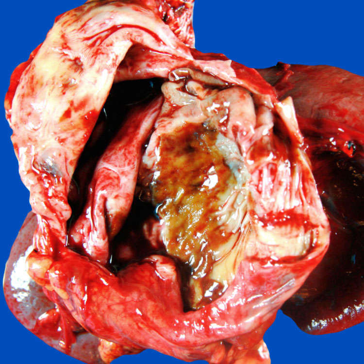
• Usually solitary and large

image Mean: 7-10 cm

• Usually multiloculated with thick walls; filled with thick, tenacious mucoid material


Microscopic


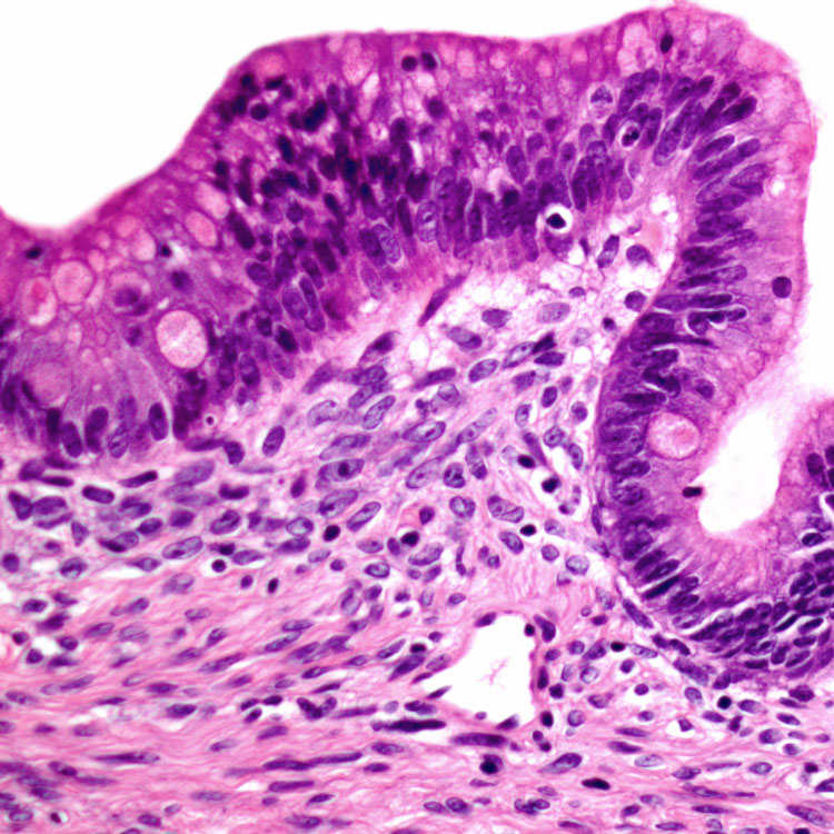
• Tall, columnar, mucin-producing epithelium with varying degrees of cellular atypia
image Invasive components can be very focal

image Recommended to submit entire lesion for microscopic evaluation

• Ovarian-type stroma is required for diagnosis
image Broad areas of stroma may be hyalinized

• Low- and high-grade dysplasia depending on degree of cytologic atypia

• Invasive carcinoma, usually pancreaticobiliary type, is present in 15% of cases

image
Mucinous Cystic Neoplasm
Unilocular cyst with a smooth, partially discolored lining is shown located in the tail of the pancreas.

image
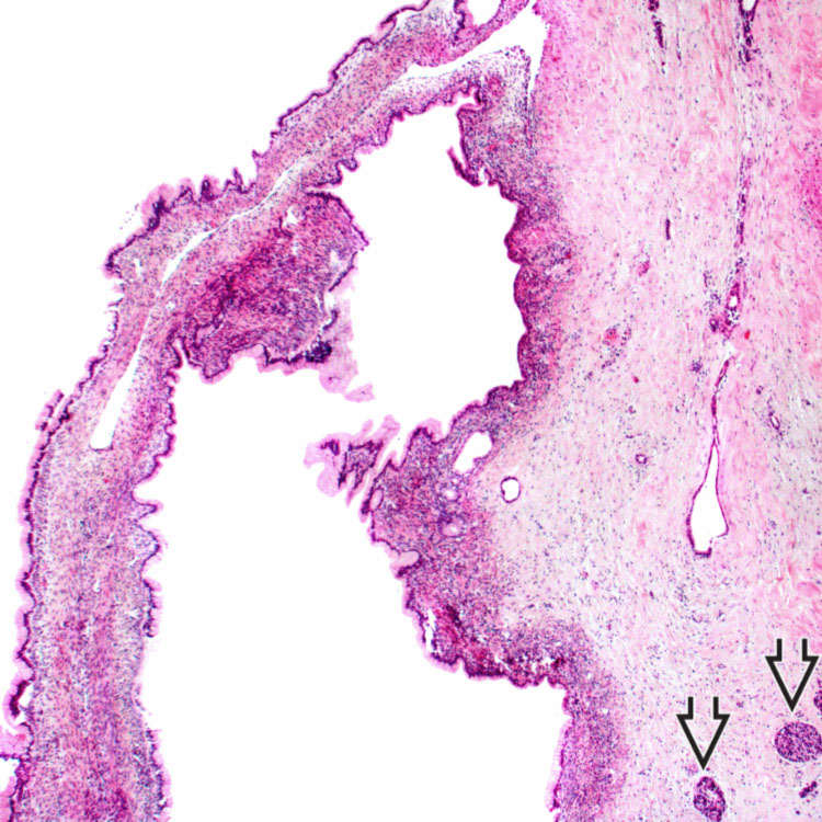
Low-Grade Dysplasia
Cyst lined by tall, columnar mucinous cells with underlying ovarian-type stroma is shown. The adjacent pancreatic parenchyma is fibrotic with a few residual islets image .

image
Intermediate-Grade Dysplasia
The lining epithelium shows enlarged, hyperchromatic nuclei with crowding and stratification.

image
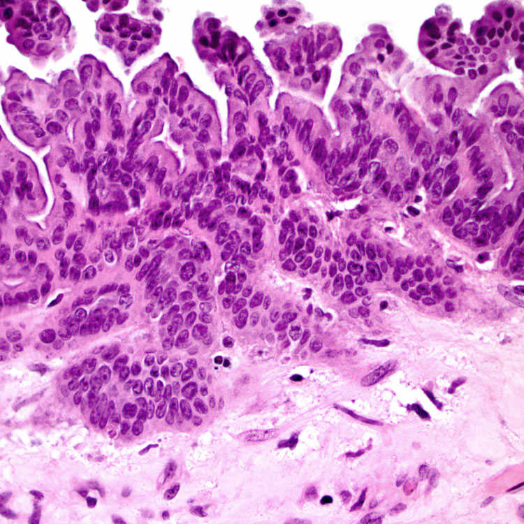
High-Grade Dysplasia
The lining epithelium shows papillary to micropapillary architecture and high-grade cytologic atypia.


TERMINOLOGY


Abbreviations


• Mucinous cystic neoplasm (MCN)


Definitions


• Neoplasm composed of mucin-producing epithelial cells associated with ovarian-type stroma


ETIOLOGY/PATHOGENESIS


Molecular Changes


• KRAS2 mutation is seen in 20% of low-grade, 33% of intermediate-grade, and 90% of high-grade MCNs

• TP53 mutation and DPC4/SMAD4 loss usually seen only in advanced MCN, in invasive component


Proposed Pathogenesis


• Ovarian-type stroma may develop from endodermal immature stroma stimulated by female hormones

• May develop from primary yolk cells implanted in pancreas during embryogenesis


CLINICAL ISSUES


Epidemiology


• Incidence
image 10% of cystic lesions in pancreas

• Age
image Range: 14-95 years (average at diagnosis: 40-50)

• Sex
image Predominantly female (F:M = 20:1)


Presentation


• Vague abdominal symptoms (pain and fullness) with compression of adjacent organs and tissues


Prognosis


• Excellent prognosis for patients with MCN without invasion
• 5-year survival rate of 50% for invasive MCN

image Size and extent of invasion (confined to pancreas vs. beyond tumor capsule) and age of patient (lower survival rate > 50 years) correlates with survival


IMAGING


CT Findings


• Usually large, well-demarcated, thick-walled, multilocular cystic mass with peripheral calcification (20%)

Only gold members can continue reading. Log In or Register to continue

Stay updated, free articles. Join our Telegram channel

Apr 20, 2017 | Posted by in PATHOLOGY & LABORATORY MEDICINE | Comments Off on Mucinous Cystic Neoplasm

Full access? Get Clinical Tree
Get Clinical Tree app for offline access