Miscellaneous Xanthomas
Jonathan B. McHugh, MD
Key Facts
Terminology
Mass-forming collection of lipidized macrophages
Reactive process usually resulting from altered serum lipid levels
Etiology/Pathogenesis
Associated with hereditary lipoproteinemias and occasionally secondary lipoproteinemias
May also occur in normolipemic patients
Clinical Issues
Usually occur in skin and subcutaneous tissue
Occasionally arise in deep soft tissues (tendon, synovium, bone)
Classified based on clinical features and gross appearance
Excellent prognosis
Microscopic Pathology
Specific classification requires clinicopathologic correlation
Generally consist of mixtures of foamy and nonfoamy macrophages with secondary changes including inflammation, fibrosis and cholesterol cleft formation
Top Differential Diagnoses
Giant cell tumor of tendon sheath
Juvenile xanthogranuloma
Plexiform xanthoma
Lipidized benign fibrous histiocytoma (dermatofibroma)
Verruciform xanthoma
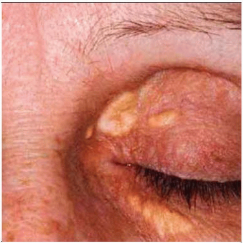 Xanthelasmas typically symmetrically involve bilateral upper and lower eyelids and periorbital skin. Sharply demarcated soft yellow papules and plaques with a yellow color are characteristic. |
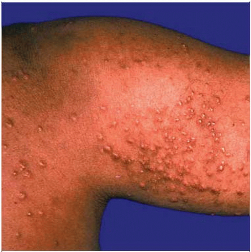 Eruptive xanthomas are characterized by the sudden appearance of crops of small yellow papules with an erythematous base. Eruptive xanthomas typically occur in the buttock, thigh, and shoulder regions. |
TERMINOLOGY
Definitions
Mass-forming collection of lipidized macrophages
Reactive process usually resulting from altered serum lipid levels
ETIOLOGY/PATHOGENESIS
Hereditary or Nonhereditary
Associated with hereditary lipoproteinemias and occasionally secondary lipoproteinemias (e.g., diabetes, hypothyroidism, primary biliary cirrhosis)
May also occur in normolipemic patients
CLINICAL ISSUES
Presentation
Usually occur in skin and subcutaneous tissue
Occasionally arise in deep soft tissues (tendon, synovium, bone)
Classified based on clinical features
Xanthelasma
Soft yellow plaques; predilection for eyelids and periorbital skin; often bilateral
Eruptive xanthoma
Sudden onset of small yellow papules with erythematous halo; predilection for gluteal region, thigh, and shoulders
Tuberous xanthoma
Firm yellow subcutaneous nodules and plaques; predilection for elbow, knee, gluteal region, and fingers
Tendinous xanthoma
Soft tissue mass associated with tendons, ligaments, &/or fascia; predilection for hands, feet, and Achilles tendon
May impair joint function but often asymptomatic
Plane xanthoma
Variably sized yellow macules; predilection for palmar creases
In normolipemic patients, consider underlying reticuloendothelial malignancy
Cerebrotendinous xanthomatosis
Rare autosomal recessive disease; sterol 27-hydroxylase gene (CYP27A) mutation
Enzyme involved in bile acid synthesis; defect results in accumulation of cholestanol, which is deposited systemically
Bilateral Achilles tendon xanthomas and cataracts; CNS symptoms include ataxia, dementia, dysarthria, psychiatric disturbances, and seizures
Treatment
May regress with medical therapy for hyperlipidemia or underlying cause if secondary
Conservative excision can be employed for large or symptomatic lesions
Prognosis
Excellent prognosis; surgically treated lesions may recur
MACROSCOPIC FEATURES
General Features
Diffuse or circumscribed with variegated yellow, tan, and white appearance
Size
Generally a few millimeters to centimeters depending on type
Tendinous xanthomas can be quite large (up to 20 cm)
MICROSCOPIC PATHOLOGY
Histologic Features
Specific classification requires clinicopathologic correlation
Consist of mixtures of foamy and nonfoamy macrophages with variable inflammation, fibrosis, and cholesterol cleft formation
Xanthelasmas and plane xanthomas consist of sheets of foamy macrophages
Eruptive xanthomas consist mostly of nonfoamy macrophages with some foamy macrophages
Tuberous and tendinous xanthomas consist of sheets of foamy macrophages with chronic inflammation, fibrosis, and cholesterol clefts with giant cells
DIFFERENTIAL DIAGNOSIS
Giant Cell Tumor of Tendon Sheath
Stay updated, free articles. Join our Telegram channel

Full access? Get Clinical Tree


