Minimally Invasive Right Hepatectomy
Shirin Sabbaghian
Allan Tsung
DEFINITION
Minimally invasive right hepatectomy is defined as resection of segments 5 to 8 using minimally invasive techniques (see Part 3, Chapter 15, FIG 2). These techniques include the following:
Pure laparoscopic
Hand assisted—The surgeon’s hand is used to assist during the laparoscopic approach.
Hybrid—The liver is mobilized laparoscopically followed by minilaparotomy to divide the liver.
Robotic—The liver is resected with robotic technique.
PATIENT HISTORY AND PHYSICAL FINDINGS
A thorough history and physical exam should be performed for each patient.
A patient’s history is important and should include careful attention to the following:
Medical history—Is the liver neoplasm benign or malignant? If the neoplasm is malignant, is it a primary of the liver or a metastasis? If the lesion is malignant, has the patient had chemotherapy? Does the patient have underlying liver disease such as cirrhosis? If cirrhosis exists, does the patient have manifestations of advanced liver disease such as esophageal varices, splenomegaly and hypersplenism, ascites, or hepatic encephalopathy? Is the patient healthy enough to withstand liver resection?
Surgical history—Has the patient had abdominal surgery in the past? If so, what type of operation and how many operations? Has the patient had prior liver or biliary tract surgery?
Social history—Alcohol use is important for consideration of concurrent liver disease.
Functional status should be considered when deciding goals of care and whether surgery is appropriate for an individual patient.
Physical exam should include evaluation for the presence of advanced liver disease such as jaundice/scleral icterus (can also be present with biliary obstruction), gynecomastia, splenomegaly, ascites, and caput medusa. Findings of advanced malignancy, including supraclavicular lymphadenopathy, should be recognized.
IMAGING AND OTHER DIAGNOSTIC STUDIES
Abdominal imaging using computed tomography (CT) or magnetic resonance imaging (MRI) within a short interval of time (within 4 to 6 weeks) prior to surgery is necessary. Attention should be focused on the number of lesions, the location of the lesions (particularly in relation to the hepatic and portal veins [PVs] as well as the inferior vena cava), the character of the background liver (normal, cirrhosis, steatosis), and the proportion of liver that will be left in situ once all hepatic disease is resected.
For CT scan, a dynamic bolus, contrast-enhanced, multidetector CT using at least a three-phase protocol is ideal. Noncontrast images of the liver are followed by rapid bolus of contrast and images are immediately obtained during peak arterial enhancement (arterial phase) as well as during PV enhancement (portal venous phase). Hypervascular tumors and tumors that receive their blood supply primarily from the hepatic artery (HA) are best visualized on arterial phase. During the portal venous phase, the liver is maximally enhanced and hypovascular lesions on a background of brightappearing parenchyma can be well-distinguished (FIG 1).
Enhanced hepatic MRI is an equivalent alternative.
Tissue sampling with core biopsy by ultrasound or CT guidance can be useful to make or confirm a diagnosis.
Additional diagnostic studies that are necessary prior to operation are basic labs, including a hepatic function panel and coagulation studies. A complete blood count including a platelet count is important to help detect advanced portal hypertension if the patient has cirrhosis. If disease-specific tumor markers are elevated, they are helpful to confirm a diagnosis as well as to follow patients during treatment.
SURGICAL MANAGEMENT AND OPERATIVE TECHNIQUE
There are several advantages of laparoscopic liver resection when compared to an open technique. These include decreased blood loss, less postoperative pain, quicker return to diet, and a shorter length of stay.1 These advantages have been demonstrated in case-controlled studies. Additionally, oncologic outcomes (margins, survival) are not compromised when laparoscopic technique is used for patients with hepatocellular carcinoma and metastatic colorectal cancer.2, 3, 4
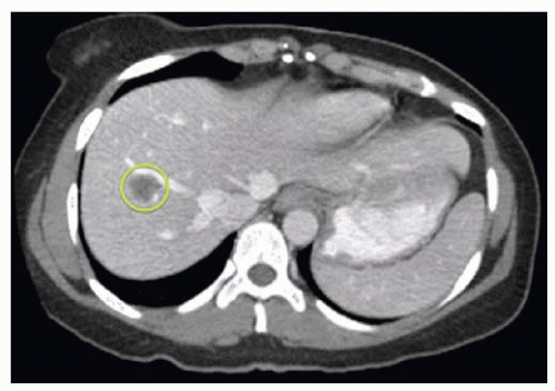
FIG 1 • CT image of liver, portal venous phase. Encircled is a metastatic colon lesion to the right liver.
During the World Consensus Conference on Laparoscopic Surgery in 2008, the international position on laparoscopic liver surgery was created—this should be used as a guide to determine which patients are eligible for minimally invasive hepatic resection.5 This statement recommends surgery with minimally invasive technique for patients with a single lesion of 5 cm or less located in segments 2 to 6. It suggests that major liver resection can be performed with minimally invasive technique but only by those experienced both with liver surgery as well as minimally invasive liver resection. Some surgeons in high-volume centers may choose to operate beyond these criteria in certain settings. Importantly, the consensus conference suggests that the surgeon should be facile with minimally invasive technique, including the skill of intracorporeal suturing should bleeding become an issue.
The decision to use hand-assisted versus pure laparoscopic technique is surgeon specific and mostly depends on comfort with either technique. As our own experience in laparoscopic hepatectomy has grown, so has our comfort with performing anatomic hepatic resection in a pure laparoscopic manner. When comparing pure laparoscopic technique to hand-assisted or hybrid technique in 113 patients undergoing anatomic liver resection, we found similar results for estimated blood loss and complications; however, interestingly, shorter operative times were noted in the pure laparoscopic group (188 minutes vs. 264 minutes for the pure vs. hand-assisted technique, P < .05).6
Consideration of the background liver is important to help decide which patients are appropriate for resection—up to 80% of a healthy liver in a relatively healthy patient can be resected without major consequence. This percentage decreases if the liver is cirrhotic.
The technique of minimally invasive right hepatectomy is, as intended, the same as for open surgery except minimally invasive equipment is used.
Two surgeons who are experienced in hepatobiliary and minimally invasive surgery are appropriate for these cases. An assistant to hold the camera is optimal, thus allowing both hands of both surgeons to be free.
TECHNIQUES
HAND-ASSISTED LAPAROSCOPIC RIGHT HEPATECTOMY
The patient is positioned supine with the arms tucked. Padded barriers are secured at the patient’s feet to prevent sliding with the anticipated use of steep reverse Trendelenburg during the case.
Access is gained to the abdominal cavity via a 5-mm port ideally in the left upper quadrant (LUQ), and pneumoperitoneum of 12 mmHg is created. Additional ports are placed using a 5-mm 30-degree scope for visualization (FIG 2). These ports include an 8-cm hand access site in the supraumbilical position. Two 12-mm ports are placed in the subxiphoid and right paramedian position, and two 5-mm ports are placed in right subcostal positions. The camera is switched to a 10-mm 30-degree scope in the right paramedian port.
Stay updated, free articles. Join our Telegram channel

Full access? Get Clinical Tree


