FIGURE 351-8 Wireless capsule endoscopy image in a patient with Crohn’s disease of the ileum shows ulcerations and narrowing of the intestinal lumen. (Courtesy of Dr. S. Reddy, Gastroenterology Division, Department of Medicine, Brigham and Women’s Hospital, Boston, Massachusetts; with permission.)
In CD, early radiographic findings in the small bowel include thickened folds and aphthous ulcerations. “Cobblestoning” from longitudinal and transverse ulcerations most frequently involves the small bowel. In more advanced disease, strictures, fistulas, inflammatory masses, and abscesses may be detected. The earliest macroscopic findings of colonic CD are aphthous ulcers. These small ulcers are often multiple and separated by normal intervening mucosa. As the disease progresses, aphthous ulcers become enlarged, deeper, and occasionally connected to one another, forming longitudinal stellate, serpiginous, and linear ulcers (see Fig. 345-4B).
The transmural inflammation of CD leads to decreased luminal diameter and limited distensibility. As ulcers progress deeper, they can lead to fistula formation. The radiographic “string sign” represents long areas of circumferential inflammation and fibrosis, resulting in long segments of luminal narrowing. The segmental nature of CD results in wide gaps of normal or dilated bowel between involved segments.
Both CT and MRI of the small bowel can be performed by enterography (CTE or MRE), using oral and IV contrast, as well as enteroclysis. Although institutional preference guides technique selection, CTE and MRE tend to be preferred over enteroclysis due to ease and patient preference. Although CTE, MRE, and small-bowel follow-through (SBFT) have been shown to be equally accurate in the identification of active small-bowel inflammation, CTE and MRE have been shown to be superior to SBFT in the detection of extraluminal complications, including fistulas, sinus tracts, and abscesses. Currently, the use of CT scans is more common than MRI due to institutional availability and expertise. However, MRI is thought to offer superior soft tissue contrast and has the added advantage of avoiding radiation exposure changes (Figs. 351-9 and 351-10). The lack of ionizing radiation is particularly appealing in younger patients and when monitoring response to therapy where serial images will be obtained. Either CTE or MRE is the first-line test for the evaluation of suspected CD and its complications. Pelvic MRI is superior to CT for demonstrating pelvic lesions such as ischiorectal abscesses and perianal fistulae (Fig. 351-11).
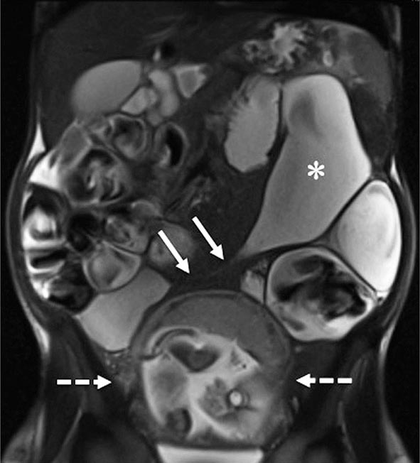
FIGURE 351-9 A coronal magnetic resonance image was obtained using a half Fourier single-shot T2-weighted acquisition with fat saturation in a 27-year-old pregnant (23 weeks’ gestation) woman. The patient had Crohn’s disease and was maintained on 6-mercaptopurine and prednisone. She presented with abdominal pain, distension, vomiting, and small-bowel obstruction. The image reveals a 7- to 10-cm long stricture at the terminal ileum (white arrows) causing obstruction and significant dilatation of the proximal small bowel (white asterisk). A fetus is seen in the uterus (dashed white arrows). (Courtesy of Drs. J. F. B. Chick and P. B. Shyn, Abdominal Imaging and Intervention, Department of Radiology, Brigham and Women’s Hospital, Harvard Medical School, Boston, Massachusetts; with permission.)
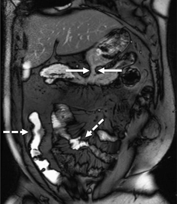
FIGURE 351-10 A coronal balanced, steady-state, free precession, T2-weighted image with fat saturation was obtained in a 32-year-old man with Crohn’s disease and prior episodes of bowel obstruction, fistulas, and abscesses. He was being treated with 6-mercaptopurine and presented with abdominal distention and diarrhea. The image demonstrates a new gastrocolic fistula (solid white arrows). Multifocal involvement of the small bowel and terminal ileum is also present (dashed white arrows). (Courtesy of Drs. J. F. B. Chick and P. B. Shyn, Abdominal Imaging and Intervention, Department of Radiology, Brigham and Women’s Hospital, Harvard Medical School, Boston, Massachusetts; with permission.)
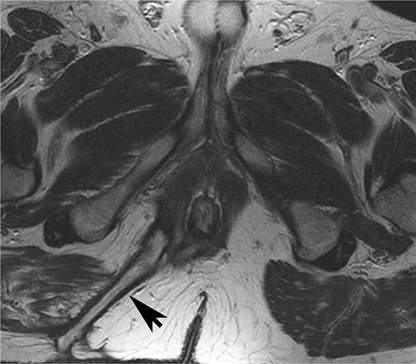
FIGURE 351-11 Axial T2-weighted magnetic resonance image obtained in a 37-year-old man with Crohn’s disease shows a linear fluid-filled perianal fistula (arrow) in the right ischioanal fossa. (Courtesy of Dr. K. Mortele, Gastrointestinal Radiology, Department of Radiology, Brigham and Women’s Hospital, Boston, Massachusetts; with permission.)
Complications Because CD is a transmural process, serosal adhesions develop that provide direct pathways for fistula formation and reduce the incidence of free perforation. Perforation occurs in 1–2% of patients, usually in the ileum but occasionally in the jejunum or as a complication of toxic megacolon. The peritonitis of free perforation, especially colonic, may be fatal. Intraabdominal and pelvic abscesses occur in 10–30% of patients with CD at some time in the course of their illness. CT-guided percutaneous drainage of the abscess is standard therapy. Despite adequate drainage, most patients need resection of the offending bowel segment. Percutaneous drainage has an especially high failure rate in abdominal wall abscesses. Systemic glucocorticoid therapy increases the risk of intraabdominal and pelvic abscesses in CD patients who have never had an operation. Other complications include intestinal obstruction in 40%, massive hemorrhage, malabsorption, and severe perianal disease.
Serologic Markers Patients with CD show a wide variation in the way they present and progress over time. Some patients present with mild disease activity and do well with generally safe and mild medications, but many others exhibit more severe disease and can develop serious complications that will require surgery. Current and developing biologic therapies can help halt progression of disease and give patients with moderate to severe CD a better quality of life. There are potential risks of biologic therapies such as infection and malignancy, and it would be optimal to determine at the time of diagnosis which patients will require more aggressive medical therapy. This same argument holds true for UC patients as well.
Subsets of patients with differing immune responses to microbial antigens have been described, and serology is often tested for perinuclear antineutrophil cytoplasmic antibodies (pANCAs) and anti-Saccharomyces cerevisiae antibodies (ASCAs). Unfortunately, these serologic markers are only marginally useful in helping to make the diagnosis of UC or CD and in predicting the course of disease. For success in diagnosing IBD and in differentiating between CD and UC, the efficacy of these serologic tests depends on the prevalence of IBD in a specific population. pANCA positivity is found in about 60–70% of UC patients and 5–10% of CD patients; 5–15% of first-degree relatives of UC patients are pANCA positive, whereas only 2–3% of the general population is pANCA positive. Sixty to 70% of CD patients, 10–15% of UC patients, and up to 5% of non-IBD controls are ASCA positive. In a patient population with a combined prevalence of UC and CD of 62%, pANCA/ASCA serology showed a sensitivity of 64% and a specificity of 94%. Positive and negative predictive values (PPVs and NPVs) for pANCA/ASCA also vary based on the prevalence of IBD in a given population. For the patient population with a prevalence of IBD of 62%, the PPV is 94%, and the NPV is 63%.
Other serologic tests include antibodies to Escherichia coli outer membrane porin protein C (OmpC), which is found in 55% of CD patients; antibodies to I2, a homologue of the bacterial transcription factor families from a Pseudomonas fluorescens–associated sequence that is found in 50–54% of CD patients; and anti-flagellin (anti-CBir1) antibodies, which have been identified in approximately 50% of CD patients.
Children with CD positive for all four immune responses (ASCA+, OmpC+, I2+, and anti-Cbir1+) may have more aggressive disease and a shorter time to progression to internal perforating and/or stricturing disease. However, larger prospective studies in both children and adults have not yet been performed and compared to CRP or other markers.
Clinical factors described at diagnosis are more helpful than serologies at predicting the natural history of CD. The initial requirements for glucocorticoid use, an age at diagnosis below 40 years and the presence of perianal disease at diagnosis, have been shown to be independently associated with subsequent disabling CD after 5 years. Except in special circumstances (such as before consideration of an ileoanal pouch anastomosis [IPAA] in a patient with indeterminate colitis), serologic markers have only minimal clinical utility.
DIFFERENTIAL DIAGNOSIS OF UC AND CD
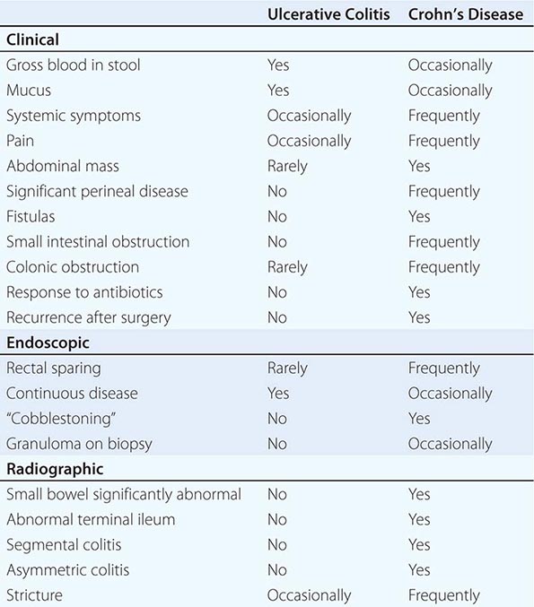
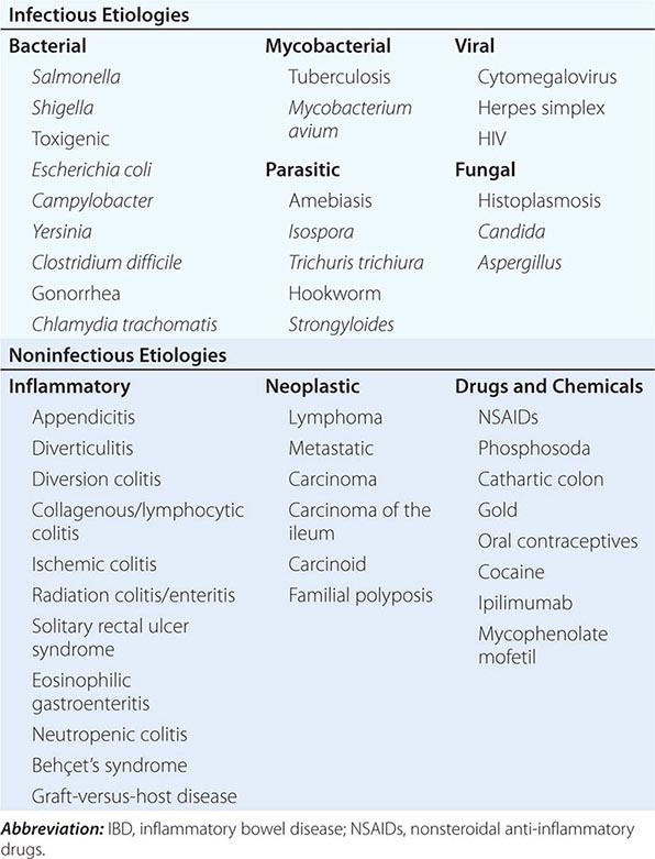
UC and CD have similar features to many other diseases. In the absence of a key diagnostic test, a combination of features is used (Table 351-5). Once a diagnosis of IBD is made, distinguishing between UC and CD is impossible initially in up to 15% of cases. These are termed indeterminate colitis. Fortunately, in most cases, the true nature of the underlying colitis becomes evident later in the course of the patient’s disease. Approximately 5% (range 1–20%) of colon resection specimens are difficult to classify as either UC or CD because they exhibit overlapping histologic features.
DIFFERENT CLINICAL, ENDOSCOPIC, AND RADIOGRAPHIC FEATURES |
INFECTIOUS DISEASES
Infections of the small intestines and colon can mimic CD or UC. They may be bacterial, fungal, viral, or protozoal in origin (Table 351-6). Campylobacter colitis can mimic the endoscopic appearance of severe UC and can cause a relapse of established UC. Salmonella can cause watery or bloody diarrhea, nausea, and vomiting. Shigellosis causes watery diarrhea, abdominal pain, and fever followed by rectal tenesmus and by the passage of blood and mucus per rectum. All three are usually self-limited, but 1% of patients infected with Salmonella become asymptomatic carriers. Yersinia enterocolitica infection occurs mainly in the terminal ileum and causes mucosal ulceration, neutrophil invasion, and thickening of the ileal wall. Other bacterial infections that may mimic IBD include C. difficile, which presents with watery diarrhea, tenesmus, nausea, and vomiting; and E. coli, three categories of which can cause colitis. These are enterohemorrhagic, enteroinvasive, and enteroadherent E. coli, all of which can cause bloody diarrhea and abdominal tenderness. Diagnosis of bacterial colitis is made by sending stool specimens for bacterial culture and C. difficile toxin analysis. Gonorrhea, Chlamydia, and syphilis can also cause proctitis.
DISEASES THAT MIMIC IBD |
GI involvement with mycobacterial infection occurs primarily in the immunosuppressed patient but may occur in patients with normal immunity. Distal ileal and cecal involvement predominates, and patients present with symptoms of small-bowel obstruction and a tender abdominal mass. The diagnosis is made most directly by colonoscopy with biopsy and culture. Mycobacterium avium-intracellulare complex infection occurs in advanced stages of HIV infection and in other immunocompromised states; it usually manifests as a systemic infection with diarrhea, abdominal pain, weight loss, fever, and malabsorption. Diagnosis is established by acid-fast smear and culture of mucosal biopsies.
Although most of the patients with viral colitis are immunosuppressed, cytomegalovirus (CMV) and herpes simplex proctitis may occur in immunocompetent individuals. CMV occurs most commonly in the esophagus, colon, and rectum but may also involve the small intestine. Symptoms include abdominal pain, bloody diarrhea, fever, and weight loss. With severe disease, necrosis and perforation can occur. Diagnosis is made by identification of characteristic intranuclear inclusions in mucosal cells on biopsy. Herpes simplex infection of the GI tract is limited to the oropharynx, anorectum, and perianal areas. Symptoms include anorectal pain, tenesmus, constipation, inguinal adenopathy, difficulty with urinary voiding, and sacral paresthesias. Diagnosis is made by rectal biopsy with identification of characteristic cellular inclusions and viral culture. HIV itself can cause diarrhea, nausea, vomiting, and anorexia. Small intestinal biopsies show partial villous atrophy; small bowel bacterial overgrowth and fat malabsorption may also be noted.
Protozoan parasites include Isospora belli, which can cause a self-limited infection in healthy hosts but causes a chronic profuse, watery diarrhea, and weight loss in AIDS patients. Entamoeba histolytica or related species infect about 10% of the world’s population; symptoms include abdominal pain, tenesmus, frequent loose stools containing blood and mucus, and abdominal tenderness. Colonoscopy reveals focal punctate ulcers with normal intervening mucosa; diagnosis is made by biopsy or serum amebic antibodies. Fulminant amebic colitis is rare but has a mortality rate of >50%.
Other parasitic infections that may mimic IBD include hookworm (Necator americanus), whipworm (Trichuris trichiura), and Strongyloides stercoralis. In severely immunocompromised patients, Candida or Aspergillus can be identified in the submucosa. Disseminated histoplasmosis can involve the ileocecal area.
NONINFECTIOUS DISEASES
Diverticulitis can be confused with CD clinically and radiographically. Both diseases cause fever, abdominal pain, tender abdominal mass, leukocytosis, elevated ESR, partial obstruction, and fistulas. Perianal disease or ileitis on small-bowel series favors the diagnosis of CD. Significant endoscopic mucosal abnormalities are more likely in CD than in diverticulitis. Endoscopic or clinical recurrence following segmental resection favors CD. Diverticular-associated colitis is similar to CD, but mucosal abnormalities are limited to the sigmoid and descending colon.
Ischemic colitis is commonly confused with IBD. The ischemic process can be chronic and diffuse, as in UC, or segmental, as in CD. Colonic inflammation due to ischemia may resolve quickly or may persist and result in transmural scarring and stricture formation. Ischemic bowel disease should be considered in the elderly following abdominal aortic aneurysm repair or when a patient has a hypercoagulable state or a severe cardiac or peripheral vascular disorder. Patients usually present with sudden onset of left lower quadrant pain, urgency to defecate, and the passage of bright red blood per rectum. Endoscopic examination often demonstrates a normal-appearing rectum and a sharp transition to an area of inflammation in the descending colon and splenic flexure.
The effects of radiotherapy on the GI tract can be difficult to distinguish from IBD. Acute symptoms can occur within 1–2 weeks of starting radiotherapy. When the rectum and sigmoid are irradiated, patients develop bloody, mucoid diarrhea and tenesmus, as in distal UC. With small-bowel involvement, diarrhea is common. Late symptoms include malabsorption and weight loss. Stricturing with obstruction and bacterial overgrowth may occur. Fistulas can penetrate the bladder, vagina, or abdominal wall. Flexible sigmoidoscopy reveals mucosal granularity, friability, numerous telangiectasias, and occasionally discrete ulcerations. Biopsy can be diagnostic.
Solitary rectal ulcer syndrome is uncommon and can be confused with IBD. It occurs in persons of all ages and may be caused by impaired evacuation and failure of relaxation of the puborectalis muscle. Single or multiple ulcerations may arise from anal sphincter overactivity, higher intrarectal pressures during defecation, and digital removal of stool. Patients complain of constipation with straining and pass blood and mucus per rectum. Other symptoms include abdominal pain, diarrhea, tenesmus, and perineal pain. Ulceration as large as 5 cm in diameter is usually seen anteriorly or anterior-laterally 3–15 cm from the anal verge. Biopsies can be diagnostic.
Several types of colitis are associated with nonsteroidal anti-inflammatory drugs (NSAIDs), including de novo colitis, reactivation of IBD, and proctitis caused by use of suppositories. Most patients with NSAID-related colitis present with diarrhea and abdominal pain, and complications include stricture, bleeding, obstruction, perforation, and fistulization. Withdrawal of these agents is crucial, and in cases of reactivated IBD, standard therapies are indicated.
There are complications of two drugs used in a hospital setting that mimic IBD. The first is ipilimumab, a drug that targets cytotoxic T lymphocyte antigen 4 (CTLA-4) and reverses T cell inhibition and is used to treat metastatic melanoma; ipilimumab has an incidence of IBD in 0.0017 cases per 100 person-years. Ipilimumab-induced colitis is typically treated with glucocorticoids or infliximab. The second is mycophenolate mofetil (MMF), an immunosuppressive agent commonly used to prevent posttransplant rejection. The colitis associated with MMF is common and can occur in more than one-third of patients taking the drug. Treatment is dose reduction or cessation of the drug.
THE ATYPICAL COLITIDES
Two atypical colitides—collagenous colitis and lymphocytic colitis—have completely normal endoscopic appearances. Collagenous colitis has two main histologic components: increased subepithelial collagen deposition and colitis with increased intraepithelial lymphocytes. The female to male ratio is 9:1, and most patients present in the sixth or seventh decades of life. The main symptom is chronic watery diarrhea. Treatments range from sulfasalazine or mesalamine and diphenoxylate/atropine (Lomotil) to bismuth to budesonide to prednisone or azathioprine/6-mercaptopurine for refractory disease. Risk factors include smoking; use of NSAIDs, proton pump inhibitors, or beta blockers; and a history of autoimmune disease.
Lymphocytic colitis has features similar to collagenous colitis, including age at onset and clinical presentation, but it has almost equal incidence in men and women and no subepithelial collagen deposition on pathologic section. However, intraepithelial lymphocytes are increased. Use of sertraline (but not beta blockers) is an additional risk factor. The frequency of celiac disease is increased in lymphocytic colitis and ranges from 9 to 27%. Celiac disease should be excluded in all patients with lymphocytic colitis, particularly if diarrhea does not respond to conventional therapy. Treatment is similar to that of collagenous colitis with the exception of a gluten-free diet for those who have celiac disease.
Diversion colitis is an inflammatory process that arises in segments of the large intestine that are excluded from the fecal stream. It usually occurs in patients with ileostomy or colostomy when a mucus fistula or a Hartmann’s pouch has been created. Clinically, patients have mucus or bloody discharge from the rectum. Erythema, granularity, friability, and, in more severe cases, ulceration can be seen on endoscopy. Histopathology shows areas of active inflammation with foci of cryptitis and crypt abscesses. Crypt architecture is normal, which differentiates it from UC. It may be impossible to distinguish from CD. Short-chain fatty acid enemas may help in diversion colitis, but the definitive therapy is surgical reanastomosis.
EXTRAINTESTINAL MANIFESTATIONS
Up to one-third of IBD patients have at least one extraintestinal disease manifestation.
DERMATOLOGIC
Erythema nodosum (EN) occurs in up to 15% of CD patients and 10% of UC patients. Attacks usually correlate with bowel activity; skin lesions develop after the onset of bowel symptoms, and patients frequently have concomitant active peripheral arthritis. The lesions of EN are hot, red, tender nodules measuring 1–5 cm in diameter and are found on the anterior surface of the lower legs, ankles, calves, thighs, and arms. Therapy is directed toward the underlying bowel disease.
Pyoderma gangrenosum (PG) is seen in 1–12% of UC patients and less commonly in Crohn’s colitis. Although it usually presents after the diagnosis of IBD, PG may occur years before the onset of bowel symptoms, run a course independent of the bowel disease, respond poorly to colectomy, and even develop years after proctocolectomy. It is usually associated with severe disease. Lesions are commonly found on the dorsal surface of the feet and legs but may occur on the arms, chest, stoma, and even the face. PG usually begins as a pustule and then spreads concentrically to rapidly undermine healthy skin. Lesions then ulcerate, with violaceous edges surrounded by a margin of erythema. Centrally, they contain necrotic tissue with blood and exudates. Lesions may be single or multiple and grow as large as 30 cm. They are sometimes very difficult to treat and often require IV antibiotics, IV glucocorticoids, dapsone, azathioprine, thalidomide, IV cyclosporine, or infliximab.
Other dermatologic manifestations include pyoderma vegetans, which occurs in intertriginous areas; pyostomatitis vegetans, which involves the mucous membranes; Sweet syndrome, a neutrophilic dermatosis; and metastatic CD, a rare disorder defined by cutaneous granuloma formation. Psoriasis affects 5–10% of patients with IBD and is unrelated to bowel activity consistent with the potential shared immunogenetic basis of these diseases. Perianal skin tags are found in 75–80% of patients with CD, especially those with colon involvement. Oral mucosal lesions, seen often in CD and rarely in UC, include aphthous stomatitis and “cobblestone” lesions of the buccal mucosa.
RHEUMATOLOGIC
Peripheral arthritis develops in 15–20% of IBD patients, is more common in CD, and worsens with exacerbations of bowel activity. It is asymmetric, polyarticular, and migratory and most often affects large joints of the upper and lower extremities. Treatment is directed at reducing bowel inflammation. In severe UC, colectomy frequently cures the arthritis.
Ankylosing spondylitis (AS) occurs in about 10% of IBD patients and is more common in CD than UC. About two-thirds of IBD patients with AS express the HLA-B27 antigen. The AS activity is not related to bowel activity and does not remit with glucocorticoids or colectomy. It most often affects the spine and pelvis, producing symptoms of diffuse low-back pain, buttock pain, and morning stiffness. The course is continuous and progressive, leading to permanent skeletal damage and deformity. Anti-TNF therapy reduces spinal inflammation and improves functional status and quality of life.
Sacroiliitis is symmetric, occurs equally in UC and CD, is often asymptomatic, does not correlate with bowel activity, and does not always progress to AS. Other rheumatic manifestations include hypertrophic osteoarthropathy, pelvic/femoral osteomyelitis, and relapsing polychondritis.
OCULAR
The incidence of ocular complications in IBD patients is 1–10%. The most common are conjunctivitis, anterior uveitis/iritis, and episcleritis. Uveitis is associated with both UC and Crohn’s colitis, may be found during periods of remission, and may develop in patients following bowel resection. Symptoms include ocular pain, photophobia, blurred vision, and headache. Prompt intervention, sometimes with systemic glucocorticoids, is required to prevent scarring and visual impairment. Episcleritis is a benign disorder that presents with symptoms of mild ocular burning. It occurs in 3–4% of IBD patients, more commonly in Crohn’s colitis, and is treated with topical glucocorticoids.
HEPATOBILIARY
Hepatic steatosis is detectable in about one-half of the abnormal liver biopsies from patients with CD and UC; patients usually present with hepatomegaly. Fatty liver usually results from a combination of chronic debilitating illness, malnutrition, and glucocorticoid therapy. Cholelithiasis occurs in 10–35% of CD patients with ileitis or ileal resection. Gallstone formation is caused by malabsorption of bile acids, resulting in depletion of the bile salt pool and the secretion of lithogenic bile.
Primary sclerosing cholangitis (PSC) is a disorder characterized by both intrahepatic and extrahepatic bile duct inflammation and fibrosis, frequently leading to biliary cirrhosis and hepatic failure; approximately 5% of patients with UC have PSC, but 50–75% of patients with PSC have IBD. PSC occurs less often in patients with CD. Although it can be recognized after the diagnosis of IBD, PSC can be detected earlier or even years after proctocolectomy. Consistent with this, the immunogenetic basis for PSC appears to be overlapping but distinct from UC based on GWAS, although both IBD and PSC are commonly pANCA positive. Most patients have no symptoms at the time of diagnosis; when symptoms are present, they consist of fatigue, jaundice, abdominal pain, fever, anorexia, and malaise. The traditional gold standard diagnostic test is endoscopic retrograde cholangiopancreatography (ERCP), but magnetic resonance cholangiopancreatography (MRCP) is also sensitive and specific. MRCP is reasonable as an initial diagnostic test in children and can visualize irregularities, multifocal strictures, and dilatations of all levels of the biliary tree. In patients with PSC, both ERCP and MRCP demonstrate multiple bile duct strictures alternating with relatively normal segments.
The bile acid ursodeoxycholic acid (ursodiol) may reduce alkaline phosphatase and serum aminotransferase levels, but histologic improvement has been marginal. High doses (25–30 mg/kg per day) may decrease the risk of colorectal dysplasia and cancer in patients with UC and PSC. Endoscopic stenting may be palliative for cholestasis secondary to bile duct obstruction. Patients with symptomatic disease develop cirrhosis and liver failure over 5–10 years and eventually require liver transplantation. PSC patients have a 10–15% lifetime risk of developing cholangiocarcinoma and then cannot be transplanted. Patients with IBD and PSC are at increased risk of colon cancer and should be surveyed yearly by colonoscopy and biopsy.
In addition, cholangiography is normal in a small percentage of patients who have a variant of PSC known as small duct primary sclerosing cholangitis. This variant (sometimes referred to as “pericholangitis”) is probably a form of PSC involving small-caliber bile ducts. It has similar biochemical and histologic features to classic PSC. It appears to have a significantly better prognosis than classic PSC, although it may evolve into classic PSC. Granulomatous hepatitis and hepatic amyloidosis are much rarer extraintestinal manifestations of IBD.
UROLOGIC
The most frequent genitourinary complications are calculi, ureteral obstruction, and ileal bladder fistulas. The highest frequency of nephrolithiasis (10–20%) occurs in patients with CD following small bowel resection. Calcium oxalate stones develop secondary to hyperoxaluria, which results from increased absorption of dietary oxalate. Normally, dietary calcium combines with luminal oxalate to form insoluble calcium oxalate, which is eliminated in the stool. In patients with ileal dysfunction, however, nonabsorbed fatty acids bind calcium and leave oxalate unbound. The unbound oxalate is then delivered to the colon, where it is readily absorbed, especially in the presence of inflammation.
METABOLIC BONE DISORDERS
Low bone mass occurs in 3–30% of IBD patients. The risk is increased by glucocorticoids, cyclosporine, methotrexate, and total parenteral nutrition (TPN). Malabsorption and inflammation mediated by IL-1, IL-6, TNF, and other inflammatory mediators also contribute to low bone density. An increased incidence of hip, spine, wrist, and rib fractures has been noted: 36% in CD and 45% in UC. The absolute risk of an osteoporotic fracture is about 1% per person per year. Fracture rates, particularly in the spine and hip, are highest among the elderly (age >60). One study noted an OR of 1.72 for vertebral fracture and an OR of 1.59 for hip fracture. The disease severity predicted the risk of a fracture. Only 13% of IBD patients who had a fracture were on any kind of antifracture treatment. Up to 20% of bone mass can be lost per year with chronic glucocorticoid use. The effect is dosage-dependent. Budesonide may also suppress the pituitary-adrenal axis and thus carries a risk of causing osteoporosis.
Osteonecrosis is characterized by death of osteocytes and adipocytes and eventual bone collapse. The pain is aggravated by motion and swelling of the joints. It affects the hips more often than knees and shoulders, and in one series, 4.3% of patients developed osteonecrosis within 6 months of starting glucocorticoids. Diagnosis is made by bone scan or MRI, and treatment consists of pain control, cord decompression, osteotomy, and joint replacement.
THROMBOEMBOLIC DISORDERS
Patients with IBD have an increased risk of both venous and arterial thrombosis even if the disease is not active. Factors responsible for the hypercoagulable state have included abnormalities of the platelet-endothelial interaction, hyperhomocysteinemia, alterations in the coagulation cascade, impaired fibrinolysis, involvement of tissue factor-bearing microvesicles, disruption of the normal coagulation system by autoantibodies, and a genetic predisposition. A spectrum of vasculitides involving small, medium, and large vessels has also been observed.
OTHER DISORDERS
More common cardiopulmonary manifestations include endocarditis, myocarditis, pleuropericarditis, and interstitial lung disease. A secondary or reactive amyloidosis can occur in patients with long-standing IBD, especially in patients with CD. Amyloid material is deposited systemically and can cause diarrhea, constipation, and renal failure. The renal disease can be successfully treated with colchicine. Pancreatitis is a rare extraintestinal manifestation of IBD and results from duodenal fistulas; ampullary CD; gallstones; PSC; drugs such as 6-mercaptopurine, azathioprine, or, very rarely, 5-ASA agents; autoimmune pancreatitis; and primary CD of the pancreas.
TREATMENT | INFLAMMATORY BOWEL DISEASE TREATMENT |
5-ASA AGENTS
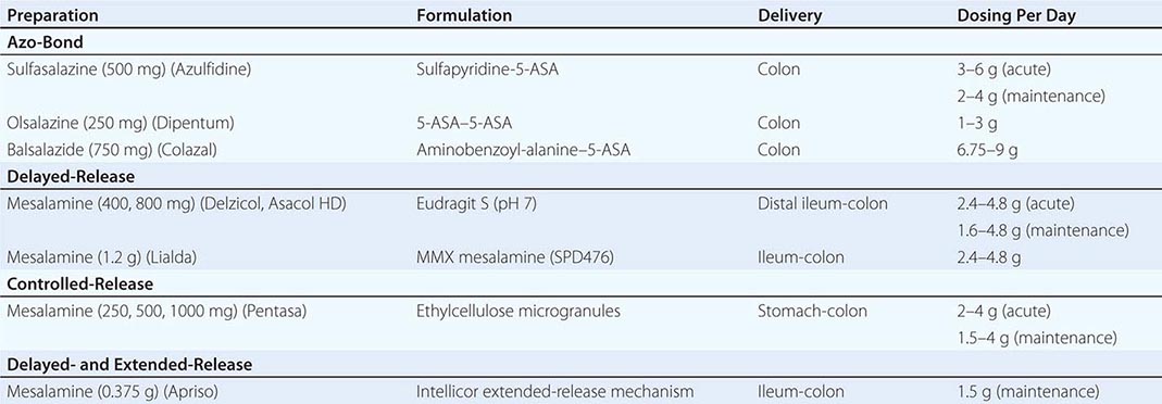
The mainstay of therapy for mild to moderate UC is sulfasalazine and the other 5-ASA agents. These agents are effective at inducing and maintaining remission in UC. They may have a limited role in inducing remission in CD but no clear role in maintenance of CD. Newer sulfa-free aminosalicylate preparations deliver increased amounts of the pharmacologically active ingredient of sulfasalazine (5-ASA, mesalamine) to the site of active bowel disease while limiting systemic toxicity. Peroxisome proliferator activated receptor γ (PPAR-γ) may mediate 5-ASA therapeutic action by decreasing nuclear localization of NF-κB. Sulfa-free aminosalicylate formulations include alternative azo-bonded carriers, 5-ASA dimers, and delayed-release and controlled-release preparations. Each has the same efficacy as sulfasalazine when equimolar concentrations are used.
Sulfasalazine was originally developed to deliver both antibacterial (sulfapyridine) and anti-inflammatory (5-ASA) therapy into the connective tissues of joints and the colonic mucosa. The molecular structure provides a convenient delivery system to the colon by allowing the intact molecule to pass through the small intestine after only partial absorption and to be broken down in the colon by bacterial azo reductases that cleave the azo bond linking the sulfa and 5-ASA moieties. Sulfasalazine is effective treatment for mild to moderate UC and is occasionally used in Crohn’s colitis, but its high rate of side effects limits its use. Although sulfasalazine is more effective at higher doses, at 6 or 8 g/d up to 30% of patients experience allergic reactions or intolerable side effects such as headache, anorexia, nausea, and vomiting that are attributable to the sulfapyridine moiety. Hypersensitivity reactions, independent of sulfapyridine levels, include rash, fever, hepatitis, agranulocytosis, hypersensitivity pneumonitis, pancreatitis, worsening of colitis, and reversible sperm abnormalities. Sulfasalazine can also impair folate absorption, and patients should be given folic acid supplements.
Balsalazide contains an azo bond binding mesalamine to the carrier molecule 4-aminobenzoyl-β-alanine; it is effective in the colon.
Olsalazine is composed of two 5-ASA radicals linked by an azo bond, which is split in the colon by bacterial reduction, and two 5-ASA molecules are released. Olsalazine is similar in effectiveness to sulfasalazine in treating UC, but up to 17% of patients experience nonbloody diarrhea caused by increased secretion of fluid in the small bowel.
Delzicol and Asacol HD (high dose) are enteric-coated forms of mesalamine with the 5-ASA being released at pH >7. They disintegrate with complete breakup of the tablet occurring in many different parts of the gut ranging from the small intestine to the splenic flexure; they have increased gastric residence when taken with a meal. Asacol has recently been discontinued and replaced with Delzicol, which lacks dibutyl phthalate (DBP), an inactive ingredient in Asacol’s enteric coating. DBP has been associated with adverse effects on the male reproductive system in animals at very high doses. Asacol HD with the same chemical in its coating is still on the market, but the human doses of DBP are within acceptable limits of toxicity.
Lialda is a once-a-day formulation of mesalamine (Multi-Matrix System [MMX]) designed to release mesalamine in the colon. The MMX technology incorporates mesalamine into a lipophilic matrix within a hydrophilic matrix encapsulated in a polymer resistant to degradation at a low pH (<7) to delay release throughout the colon. The safety profile appears to be comparable to other 5-ASA formulations.
Apriso is a formulation containing encapsulated mesalamine granules that delivers mesalamine to the terminal ileum and colon via a proprietary extended-release mechanism (Intellicor). The outer coating (Eudragit L) dissolves at a pH >6. In addition, there is a polymer matrix core that aids in sustained release throughout the colon. Because Lialda and Apriso are given once daily, an anticipated benefit is improved compliance compared with two to four daily doses required for other mesalamine preparations.
Pentasa is another mesalamine formulation that uses an ethylcellulose coating to allow water absorption into small beads containing the mesalamine. Water dissolves the 5-ASA, which then diffuses out of the bead into the lumen. Disintegration of the capsule occurs in the stomach. The microspheres then disperse throughout the entire GI tract from the small intestine through the distal colon in both fasted and fed conditions.
Salofalk® Granu-Stix, an unencapsulated version of mesalamine, has been in use in Europe for induction and maintenance of remission for several years.
Appropriate doses of the 5-ASA compounds are shown in Table 351-7. Some 50–75% of patients with mild to moderate UC improve when treated with 5-ASA doses equivalent to 2 g/d of mesalamine; the dose response continues up to at least 4.8 g/d. As a general rule, 5-ASA agents act within 2–4 weeks. 5-ASA doses equivalent to 1.5–4 g/d of mesalamine maintain remission in 50–75% of patients with UC.
ORAL 5-ASA PREPARATIONS |
More common side effects of the 5-ASA medications include headaches, nausea, hair loss, and abdominal pain. Rare side effects of the 5-ASA medications include renal impairment, hematuria, pancreatitis, and paradoxical worsening of colitis. Renal function tests and urinalysis should be checked yearly.
Topical Rowasa enemas are composed of mesalamine and are effective in mild-to-moderate distal UC. Clinical response occurs in up to 80% of UC patients with colitis distal to the splenic flexure. Combination therapy with mesalamine in both oral and enema form is more effective than either treatment alone for both distal and extensive UC.
Canasa suppositories composed of mesalamine are effective in treating proctitis.
GLUCOCORTICOIDS
The majority of patients with moderate to severe UC benefit from oral or parenteral glucocorticoids. Prednisone is usually started at doses of 40–60 mg/d for active UC that is unresponsive to 5-ASA therapy. Parenteral glucocorticoids may be administered as hydrocortisone, 300 mg/d, or methylprednisolone, 40–60 mg/d. A new glucocorticoid for UC, budesonide (Uceris), is released entirely in the colon and has minimal to no glucocorticoid side effects. The dose is 9 mg/d for 8 weeks, and no taper is required. Topically applied glucocorticoids are also beneficial for distal colitis and may serve as an adjunct in those who have rectal involvement plus more proximal disease. Hydrocortisone enemas or foam may control active disease, although they have no proven role as maintenance therapy. These glucocorticoids are significantly absorbed from the rectum and can lead to adrenal suppression with prolonged administration. Topical 5-ASA therapy is more effective than topical steroid therapy in the treatment of distal UC.
Glucocorticoids are also effective for treatment of moderate to severe CD and induce a 60–70% remission rate compared to a 30% placebo response. The systemic effects of standard glucocorticoid formulations have led to the development of more potent formulations that are less well-absorbed and have increased first-pass metabolism. Controlled ileal-release budesonide has been nearly equal to prednisone for ileocolonic CD with fewer glucocorticoid side effects. Budesonide is used for 2–3 months at a dose of 9 mg/d, and then tapered. Budesonide 6 mg/d is effective in reducing relapse rates at 3–6 months but not at 12 months in CD patients with a medically induced remission.
Glucocorticoids play no role in maintenance therapy in either UC or CD. Once clinical remission has been induced, they should be tapered according to the clinical activity, normally at a rate of no more than 5 mg/week. They can usually be tapered to 20 mg/d within 4–5 weeks but often take several months to be discontinued altogether. The side effects are numerous, including fluid retention, abdominal striae, fat redistribution, hyperglycemia, subcapsular cataracts, osteonecrosis, osteoporosis, myopathy, emotional disturbances, and withdrawal symptoms. Most of these side effects, aside from osteonecrosis, are related to the dose and duration of therapy.
ANTIBIOTICS
Antibiotics have no role in the treatment of active or quiescent UC. However, pouchitis, which occurs in about a third of UC patients after colectomy and IPAA, usually responds to treatment with metronidazole and/or ciprofloxacin.
Metronidazole is effective in active inflammatory, fistulous, and perianal CD and may prevent recurrence after ileal resection. The most effective dose is 15–20 mg/kg per day in three divided doses; it is usually continued for several months. Common side effects include nausea, metallic taste, and disulfiram-like reaction. Peripheral neuropathy can occur with prolonged administration (several months) and on rare occasions is permanent despite discontinuation. Ciprofloxacin (500 mg bid) is also beneficial for inflammatory, perianal, and fistulous CD but has been associated with Achilles tendinitis and rupture. Both ciprofloxacin and metronidazole antibiotics can be used as first-line drugs for short periods of time in active inflammatory, fistulizing, and perianal CD.
AZATHIOPRINE AND 6-MERCAPTOPURINE
Azathioprine and 6-mercaptopurine (6-MP) are purine analogues commonly employed in the management of glucocorticoid-dependent IBD. Azathioprine is rapidly absorbed and converted to 6-MP, which is then metabolized to the active end product, thioinosinic acid, an inhibitor of purine ribonucleotide synthesis and cell proliferation. These agents also inhibit the immune response. Efficacy can be seen as early as 3–4 weeks but can take up to 4–6 months. Adherence can be monitored by measuring the levels of 6-thioguanine and 6-methyl-mercaptopurine, end products of 6-MP metabolism. Azathioprine (2–3 mg/kg per day) and 6-MP (1–1.5 mg/kg per day) have been used successfully as glucocorticoid-sparing agents in up to two-thirds of UC and CD patients previously unable to be weaned from glucocorticoids. They are also used as maintenance therapy in UC and CD and for treating active perianal disease and fistulas in CD. In addition, 6-MP or azathioprine is effective for postoperative prophylaxis of CD.
Although azathioprine and 6-MP are usually well tolerated, pancreatitis occurs in 3–4% of patients, typically presents within the first few weeks of therapy, and is completely reversible when the drug is stopped. Other side effects include nausea, fever, rash, and hepatitis. Bone marrow suppression (particularly leukopenia) is dose-related and often delayed, necessitating regular monitoring of the complete blood cell count (CBC). Additionally, 1 in 300 individuals lacks thiopurine methyltransferase, the enzyme responsible for drug metabolism to inactive end-products (6-methylmercaptopurine); an additional 11% of the population are heterozygotes with intermediate enzyme activity. Both are at increased risk of toxicity because of increased accumulation of active 6-thioguanine metabolites. Although 6-thioguanine and 6-methylmercaptopurine levels can be followed to determine correct drug dosing and reduce toxicity, weight-based dosing is an acceptable alternative. CBCs and liver function tests should be monitored frequently regardless of dosing strategy. IBD patients treated with azathioprine/6-MP are at approximately a fourfold increased risk of developing a lymphoma. This increased risk could be a result of the medications, the underlying disease, or both.
METHOTREXATE
Methotrexate (MTX) inhibits dihydrofolate reductase, resulting in impaired DNA synthesis. Additional anti-inflammatory properties may be related to decreased IL-1 production. Intramuscular (IM) or subcutaneous (SC) MTX (25 mg/week) is effective in inducing remission and reducing glucocorticoid dosage; 15 mg/week is effective in maintaining remission in active CD. Potential toxicities include leukopenia and hepatic fibrosis, necessitating periodic evaluation of CBCs and liver enzymes. The role of liver biopsy in patients on long-term MTX is uncertain but is probably limited to those with increased liver enzymes. Hypersensitivity pneumonitis is a rare but serious complication of therapy.
CYCLOSPORINE
Cyclosporine (CSA) is a lipophilic peptide with inhibitory effects on both the cellular and humoral immune systems. CSA blocks the production of IL-2 by T helper lymphocytes. CSA binds to cyclophilin, and this complex inhibits calcineurin, a cytoplasmic phosphatase enzyme involved in the activation of T cells. CSA also indirectly inhibits B cell function by blocking helper T cells. CSA has a more rapid onset of action than 6-MP and azathioprine.
CSA is most effective when given at 2–4 mg/kg per day IV in severe UC that is refractory to IV glucocorticoids, with 82% of patients responding. CSA can be an alternative to colectomy. The long-term success of oral CSA is not as dramatic, but if patients are started on 6-MP or azathioprine at the time of hospital discharge, remission can be maintained. For the 2 mg/kg dose, levels as measured by monoclonal radioimmunoassay or by the high-performance liquid chromatography assay should be maintained between 150 and 350 ng/mL.
CSA may cause significant toxicity; renal function should be monitored frequently. Hypertension, gingival hyperplasia, hypertrichosis, paresthesias, tremors, headaches, and electrolyte abnormalities are common side effects. Creatinine elevation calls for dose reduction or discontinuation. Seizures may also complicate therapy, especially if the patient is hypomagnesemic or if serum cholesterol levels are <3.1 mmol/L (<120 mg/dL). Opportunistic infections, most notably Pneumocystis carinii pneumonia, may occur with combination immunosuppressive treatment; prophylaxis should be given. Major adverse events occurred in 15% of patients in one large study, including nephrotoxicity not responding to dose adjustment, serious infections, seizures, anaphylaxis, and death of two patients. This high incidence suggests that vigorous monitoring by experienced clinicians at tertiary care centers may be required. To compare IV cyclosporine versus infliximab, a large trial was conducted in Europe by the GETAID group. The results indicated identical 7-day response rates between cyclosporine 2 mg/kg (with doses adjusted for levels of 150–250 ng/mL) and infliximab 5 mg/kg, with both groups achieving response rates of 85%. Serious infections occurred in 5 of 55 cyclosporine patients and 4 of 56 infliximab patients. Response rates were similar in the two groups at day 98 among patients treated with oral cyclosporine versus infliximab at the usual induction dose and maintenance dose regimen (40% and 46%, respectively). In light of data showing equal efficacy of CSA and infliximab in severe UC, more physicians are relying on infliximab rather than CSA in these patients.
TACROLIMUS
Tacrolimus is a macrolide antibiotic with immunomodulatory properties similar to CSA. It is 100 times as potent as CSA and is not dependent on bile or mucosal integrity for absorption. These pharmacologic properties enable tacrolimus to have good oral absorption despite proximal small bowel Crohn’s involvement. It has shown efficacy in children with refractory IBD and in adults with extensive involvement of the small bowel. It is also effective in adults with glucocorticoid-dependent or refractory UC and CD as well as refractory fistulizing CD.
BIOLOGIC THERAPIES
Biologic therapy was traditionally reserved for moderately to severely ill patients with CD who had failed other therapies. However, it is now commonly given as an initial therapy for patients with moderate to severe CD in order to prevent future disease complications. Patients who respond to biologic therapies enjoy an improvement in clinical symptoms; a better quality of life; less disability, fatigue, and depression; and fewer surgeries and hospitalizations.
Anti-TNF Therapies The first biologic therapy approved for CD was infliximab, a chimeric IgG1 antibody against TNF-α, which is now also approved for treatment of moderately to severely active UC. Of active CD patients refractory to glucocorticoids, 6-MP, or 5-ASA, 65% will respond to IV infliximab (5 mg/kg); one-third will enter complete remission. The ACCENT I (A Crohn’s Disease Clinical Trial Evaluating Infliximab in a New Long-Term Treatment Regimen) study showed that of the patients who experience an initial response, 40% will maintain remission for at least 1 year with repeated infusions of infliximab every 8 weeks.
Infliximab is also effective in CD patients with refractory perianal and enterocutaneous fistulas, with the ACCENT II trial showing a 68% response rate (50% reduction in fistula drainage) and a 50% complete remission rate. Reinfusion, typically every 8 weeks, is necessary to continue therapeutic benefits in many patients.
The SONIC (Study of Biologic and Immunomodulator-Naive Patients with Crohn’s Disease) trial compared infliximab plus azathioprine, infliximab alone, and azathioprine alone in immunomodulator- and biologic-naive patients with moderate to severe CD. At 1 year, the infliximab plus azathioprine group had a glucocorticoid-free remission rate of 46% compared with 35% for infliximab alone and 24% for azathioprine alone. There was also complete mucosal healing at week 26 with the combined approach relative to either infliximab or azathioprine alone (44% vs 30% vs 17%). The adverse events were equal between groups.
Two large trials of infliximab in moderate to severe UC also showed efficacy with a response rate of 37–49%, with about one-fifth of patients maintaining remission after 54 weeks. Dosing for UC and CD are identical, with induction dosing at 0, 2, and 6 weeks and every 8 weeks thereafter. There is a similar study to SONIC in patients with moderate to severe UC. After 16 weeks of therapy, UC patients taking azathioprine plus infliximab had a glucocorticoid-free remission rate of 40% compared to 24% (article now published) and 22% of those on azathioprine and infliximab alone, respectively. This is even further evidence for “top-down” or more aggressive therapy for both moderate to severe CD and UC.
Adalimumab is a recombinant human monoclonal IgG1 antibody containing only human peptide sequences and is injected subcutaneously. Adalimumab binds TNF and neutralizes its function by blocking the interaction between TNF and its cell-surface receptor. Therefore, it seems to have a similar mechanism of action to infliximab but with less immunogenicity. Adalimumab has been approved for treatment of moderate to severe CD. CHARM (Crohn’s Trial of the Fully Human Adalimumab for Remission Maintenance) is an adalimumab maintenance study in patients who responded to adalimumab induction therapy. About 50% of the patients in this trial were previously treated with infliximab. Remission rates ranged from 42–48% of infliximab-naïve patients at 1 year compared with remission rates of 31–34% in patients who had previously received infliximab. Another trial showed a remission rate of 21% at 4 weeks in patients who had initially responded to and then failed infliximab. In clinical practice, the remission rate in patients taking adalimumab increases with a dose increase to 40 mg weekly instead of every other week. Adalimumab is now also approved for the treatment of moderately to severely active UC.
Certolizumab pegol is a pegylated form of an anti-TNF Fab portion of an antibody administered SC once monthly. SC certolizumab pegol was effective for induction of clinical response in patients with active inflammatory CD. In the PRECISE II (Pegylated Antibody Fragment Evaluation in Crohn’s Disease) trial of maintenance therapy with certolizumab in patients who responded to certolizumab induction, the results were similar to the CHARM trial. At week 26, the subgroup of patients who were infliximab naïve had a response of 69% as compared to 44% in patients who had previously received infliximab.
Golimumab is another fully human IgG1 antibody against TNF-α and is currently approved for the treatment of moderately to severely active UC. All of the patients in the golimumab trial were infliximab-naive. Like adalimumab and certolizumab, golimumab is injected SC.
Side Effects of Anti-TNF Therapies • DEVELOPMENT OF ANTIBODIES The development of antibodies to infliximab (ATIs) is associated with an increased risk of infusion reactions and a decreased response to treatment. Current practice does not include giving on-demand or episodic infusions in contrast to periodic (every 8 week) infusions because patients are most likely to develop ATIs. ATIs are generally present when the quality of response or the response duration to infliximab infusion decreases. Decreasing the dosing intervals or increasing the dosage to 10 mg/kg may restore the efficacy. There are commercial assays for both infliximab and adalimumab antibodies and trough levels to determine optimal dosing. If a patient has high ATIs and a low trough level of infliximab, it is best to switch to another anti-TNF therapy. Most acute infusion reactions and serum sickness can be managed with glucocorticoids and antihistamines. Some reactions can be serious and would necessitate a change in therapy, especially if a patient has ATIs.
NON-HODGKIN’S LYMPHOMA (NHL) The baseline risk of NHL in CD patients is 2:10,000, which is slightly higher than in the general population. Azathioprine and/or 6-MP therapy increases the risk to about 4:10,000. The highest risk for thiopurine-associated NHL is in patients over 65 years old, with a moderate risk in those between the ages of 50 and 65. Anti-TNF therapy increases the risk to approximately 6:10,000.
HEPATOSPLENIC T CELL LYMPHOMA (HSTCL) HSTCL is a nearly universally fatal lymphoma in patients with or without CD. In patients with CD, events reported to the Food and Drug Administration Adverse Event Reporting System (FDA AERS) and search of PubMed and Embase published case reports demonstrate a total of 37 unique cases. Eighty-six percent of the patients were male, with a median age of 26 years. Patients had CD for a mean of 10 years before the diagnosis of HSTCL. Thirty-six cases had used either 6-MP or azathioprine, and 28 cases had used infliximab. Of these 28 cases, 27 had also used 6-MP or azathioprine. The other case had a history of both infliximab and adalimumab exposure.
SKIN LESIONS New-onset psoriasiform skin lesions develop in nearly 5% of IBD patients treated with anti-TNF therapy. Most often, these can be treated topically, and rarely, anti-TNF therapy must be decreased, switched, or stopped. The risk of melanoma is increased almost twofold with anti-TNF and not thiopurine use. The risk of nonmelanoma skin cancer is increased with thiopurines and biologics, especially with 1 year of follow-up or greater. Patients on these medications should have a skin check at least once a year.
INFECTIONS All of the anti-TNF drugs are associated with an increased risk of infections, particularly reactivation of latent tuberculosis and opportunistic fungal infections including disseminated histoplasmosis and coccidioidomycosis. It is recommended that patients have a purified protein derivative (PPD) or a QuantiFERON-TB gold test as well as a chest x-ray before initiation of anti-TNF therapy. Patients over 65 have a higher rate of infections and death on infliximab or adalimumab than those younger than 65 years of age.
OTHER Acute liver injury due to reactivation of hepatitis B virus and to autoimmune effects and cholestasis has been reported. Rarely, infliximab and the other anti-TNF drugs have been associated with optic neuritis, seizures, new onset or exacerbation of clinical symptoms, and radiographic evidence of central nervous system demyelinating disorders, including multiple sclerosis. They may exacerbate symptoms in patients with New York Heart Association functional class III/IV heart failure.
ANTI-INTEGRINS Integrins are expressed on the cell surface of leukocytes and serve as mediators of leukocyte adhesion to vascular endothelium. α4-Integrin along with its β1 or β7 subunit interact with endothelial ligands termed adhesion molecules. Interaction between α4β7 and mucosal addressin cellular adhesion molecule (MAdCAM-1) is important in lymphocyte trafficking to gut mucosa.
Natalizumab is a recombinant humanized IgG4 antibody against α4-integrin that has been shown to be effective in induction and maintenance of patients with CD. It has been approved since February 2008 for the treatment of patients with CD refractory or intolerant to anti-TNF therapy. The rates of response and remission at 3 months are about 60% and 40%, respectively, with a sustained remission rate of about 40% at 36 weeks.
One case of progressive multifocal leukoencephalopathy (PML) after eight infusions of natalizumab was observed among 1043 patients in the clinical trials for CD, and two patients developed PML in the multiple sclerosis (MS) trials after a median of 120 weeks. There were 410 postmarketing cases of PML, 408 in MS and 2 in CD. The most important risk factor for development of PML is exposure to the John Cunningham (JC) polyomavirus, seen in 50–55% of the adult population. The other two risk factors for development of PML are longer duration of treatment, especially beyond 2 years, and prior treatment with an immunosuppressant medication. Patients with all three risk factors have an estimated risk of 11:1000.
The FDA approved a commercial enzyme-linked immunosorbent assay (ELISA) kit to assay anti-JC viral antibodies (Stratify JCV Antibody ELISA; Focus Diagnostics, Cypress, CA) in early 2012. The test is 99% accurate in stratifying risk of PML. It is recommended that all patients be tested prior to initiating natalizumab therapy. JC virus serologies are then measured every 6 months because 1–2% of patients will seroconvert yearly. All patients taking natalizumab and their providers must be enrolled in the TOUCH (Tysabri Outreach Unified Commitment for Health) pharmacovigilance program. Natalizumab is administered IV, 300 mg every 4 weeks. Labeling requirements mandate that it not be used in combination with any immunosuppressant medications.
Vedolizumab, another leukocyte trafficking inhibitor, is indicated for patients who have had an inadequate response or lost response to, or were intolerant of a TNF blocker or immunomodulator; or had an inadequate response or were intolerant to, or demonstrated dependence on glucocorticoids. It is an option for patients who are JC antibody positive since it does not cross the blood-brain barrier. Vedolizumab is a monoclonal antibody directed against α4β7 integrin specifically and has the ability to convey gut-selective immunosuppression.
THERAPIES IN DEVELOPMENT
Ustekinumab, a fully human IgG1 monoclonal antibody, blocks the biologic activity of IL-12 and IL-23 through their common p40 subunit by inhibiting the interaction of these cytokines with their receptors on T cells, natural killer cells, and antigen presenting cells. It shows efficacy in moderate to severe CD in clinical trials.
Tofacitinib is an oral inhibitor of Janus kinases 1, 3, and, to a lesser extent, 2. It is expected to block signaling involving common gamma chain–containing cytokines including IL-2, IL-4, IL-7, IL-9, IL-15, and IL-21. These cytokines are integral to lymphocyte activation, function, and proliferation. It is effective in moderate to severe UC in clinical trials.
NUTRITIONAL THERAPIES
Dietary antigens may stimulate the mucosal immune response. Patients with active CD respond to bowel rest, along with TPN. Bowel rest and TPN are as effective as glucocorticoids at inducing remission of active CD but are not effective as maintenance therapy. Enteral nutrition in the form of elemental or peptide-based preparations is also as effective as glucocorticoids or TPN, but these diets are not palatable. Enteral diets may provide the small intestine with nutrients vital to cell growth and do not have the complications of TPN. In contrast to CD, dietary intervention does not reduce inflammation in UC. Standard medical management of UC and CD is shown in Fig. 351-12.
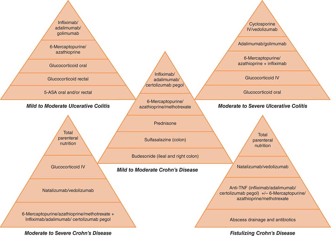
FIGURE 351-12 Medical management of inflammatory bowel disease. 5-ASA, 5-aminosalicylic acid; CD, Crohn’s disease; UC, ulcerative colitis.
SURGICAL THERAPY
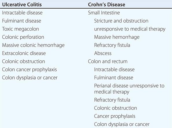
Ulcerative Colitis Nearly one-half of patients with extensive chronic UC undergo surgery within the first 10 years of their illness. The indications for surgery are listed in Table 351-8. Morbidity is about 20% for elective, 30% for urgent, and 40% for emergency proctocolectomy. The risks are primarily hemorrhage, contamination and sepsis, and neural injury. The operation of choice is an ileoanal J pouch anastomosis (IPAA).
INDICATIONS FOR SURGERY |
Because UC is a mucosal disease, the rectal mucosa can be dissected and removed down to the dentate line of the anus or about 2 cm proximal to this landmark. The ileum is fashioned into a pouch that serves as a neorectum. This ileal pouch is then sutured circumferentially to the anus in an end-to-end fashion. If performed carefully, this operation preserves the anal sphincter and maintains continence. The overall operative morbidity is 10%, with the major complication being bowel obstruction. Pouch failure necessitating conversion to permanent ileostomy occurs in 5–10% of patients. Some inflamed rectal mucosa is usually left behind, and thus endoscopic surveillance is necessary. Primary dysplasia of the ileal mucosa of the pouch has occurred rarely.
Patients with IPAA usually have about 6–10 bowel movements a day. On validated quality-of-life indices, they report better performance in sports and sexual activities than ileostomy patients. The most frequent complication of IPAA is pouchitis in about 30–50% of patients with UC. This syndrome consists of increased stool frequency, watery stools, cramping, urgency, nocturnal leakage of stool, arthralgias, malaise, and fever. Pouch biopsies may distinguish true pouchitis from underlying CD. Although pouchitis usually responds to antibiotics, 3–5% of patients remain refractory and may require glucocorticoids, immunomodulators, anti-TNF therapy, or even pouch removal. A highly concentrated probiotic preparation with four strains of Lactobacillus, three strains of Bifidobacterium, and one strain of Streptococcus salivarius can prevent the recurrence of pouchitis when taken daily.
Crohn’s Disease Most patients with CD require at least one operation in their lifetime. The need for surgery is related to duration of disease and the site of involvement. Patients with small-bowel disease have an 80% chance of requiring surgery. Those with colitis alone have a 50% chance. Surgery is an option only when medical treatment has failed or complications dictate its necessity. The indications for surgery are shown in Table 351-8.
SMALL INTESTINAL DISEASE Because CD is chronic and recurrent, with no clear surgical cure, as little intestine as possible is resected. Current surgical alternatives for treatment of obstructing CD include resection of the diseased segment and strictureplasty. Surgical resection of the diseased segment is the most frequently performed operation, and in most cases, primary anastomosis can be done to restore continuity. If much of the small bowel has already been resected and the strictures are short, with intervening areas of normal mucosa, strictureplasties should be done to avoid a functionally insufficient length of bowel. The strictured area of intestine is incised longitudinally and the incision sutured transversely, thus widening the narrowed area. Complications of strictureplasty include prolonged ileus, hemorrhage, fistula, abscess, leak, and restricture.
There is evidence that mesalamine, nitroimidazole antibiotics, 6-MP/azathioprine, infliximab, and adalimumab are all superior to placebo for the prevention of postoperative recurrence of CD. Mesalamine is the least effective, and the side effects of the nitroimidazole antibiotics limit their use. Risk factors for early recurrence of disease include cigarette smoking, penetrating disease (internal fistulas, abscesses, or other evidence of penetration through the wall of the bowel), early recurrence since a previous surgery, multiple surgeries, and a young age at the time of the first surgery. Aggressive postoperative treatment with 6-MP/azathioprine, infliximab, or adalimumab should be considered for this group of patients. It is also recommended to evaluate for endoscopic recurrence of CD via a colonoscopy, if possible, 6 months after surgery.
COLORECTAL DISEASE A greater percentage of patients with Crohn’s colitis require surgery for intractability, fulminant disease, and anorectal disease. Several alternatives are available, ranging from the use of a temporary loop ileostomy to resection of segments of diseased colon or even the entire colon and rectum. For patients with segmental involvement, segmental colon resection with primary anastomosis can be performed. In 20–25% of patients with extensive colitis, the rectum is spared sufficiently to consider rectal preservation. Most surgeons believe that an IPAA is contraindicated in CD due to the high incidence of pouch failure. A diverting colostomy may help heal severe perianal disease or rectovaginal fistulas, but disease almost always recurs with reanastomosis. These patients often require a total proctocolectomy and ileostomy.
INFLAMMATORY BOWEL DISEASE AND PREGNANCY
Patients with quiescent UC and CD have normal fertility rates; the fallopian tubes can be scarred by the inflammatory process of CD, especially on the right side because of the proximity of the terminal ileum. In addition, perirectal, perineal, and rectovaginal abscesses and fistulae can result in dyspareunia. Infertility in men can be caused by sulfasalazine but reverses when treatment is stopped. In women who have an ileoanal J pouch anastomosis, most studies show that the fertility rate is reduced to about 50–80% of normal. This is due to scarring or occlusion of the fallopian tubes secondary to pelvic inflammation.
In mild or quiescent UC and CD, fetal outcome is nearly normal. Spontaneous abortions, stillbirths, and developmental defects are increased with increased disease activity, not medications. The courses of CD and UC during pregnancy mostly correlate with disease activity at the time of conception. Patients should be in remission for 6 months before conceiving. Most CD patients can deliver vaginally, but cesarean delivery may be the preferred route of delivery for patients with anorectal and perirectal abscesses and fistulas to reduce the likelihood of fistulas developing or extending into the episiotomy scar. Unless they desire multiple children, UC patients with an IPAA should consider a cesarean delivery due to an increased risk of future fecal incontinence.
Sulfasalazine, Lialda, Apriso, Delzicol, and balsalazide are safe for use in pregnancy and nursing with the caveat that additional folate supplementation must be given with sulfasalazine. Asacol HD and olsalazine are considered by the FDA to be class C agents in pregnancy and thus not recommended. Topical 5-ASA agents are also safe during pregnancy and nursing. Glucocorticoids are generally safe for use during pregnancy and are indicated for patients with moderate to severe disease activity. The amount of glucocorticoids received by the nursing infant is minimal. The safest antibiotics to use for CD in pregnancy for short periods of time (weeks, not months) are ampicillin and cephalosporins. Metronidazole can be used in the second or third trimester. Ciprofloxacin causes cartilage lesions in immature animals and should be avoided because of the absence of data on its effects on growth and development in humans.
6-MP and azathioprine pose minimal or no risk during pregnancy, but experience is limited. If the patient cannot be weaned from the drug or has an exacerbation that requires 6-MP/azathioprine during pregnancy, she should continue the drug with informed consent. Breast milk has been shown to contain negligible levels of 6-MP/azathioprine when measured in a limited number of patients.
Little data exist on CSA in pregnancy. In a small number of patients with severe IBD treated with IV CSA during pregnancy, 80% of pregnancies were successfully completed without development of renal toxicity or congenital malformations. However, because of the lack of data, CSA should probably be avoided unless the patient would otherwise require surgery.
MTX is contraindicated in pregnancy and nursing. In a large prospective study, no increased risk of stillbirths, miscarriages, or spontaneous abortions was seen with infliximab, adalimumab, or certolizumab, which are all class B drugs. Infliximab and adalimumab are IgG1 antibodies and are actively transported across the placenta in the late second and third trimester. Infants can have serum levels of both infliximab and adalimumab up to 7 months of age, and live vaccines should be avoided during this time. Certolizumab crosses the placenta by passive diffusion, and infant serum and cord blood levels are minimal. The anti-TNF drugs are relatively safe in nursing. Miniscule levels of both infliximab and adalimumab, but not certolizumab, have been reported in breast milk. These levels are of no clinical significance. It is recommended that drugs not be switched during pregnancy unless necessitated by the medical condition of the IBD. Natalizumab is considered as a class C drug because there is limited data in pregnancy.
Surgery in UC should be performed only for emergency indications, including severe hemorrhage, perforation, and megacolon refractory to medical therapy. Total colectomy and ileostomy carry a 50% risk of postoperative spontaneous abortion. Fetal mortality is also high in CD requiring surgery. Patients with IPAAs have increased nighttime stool frequency during pregnancy that resolves postpartum. Transient small-bowel obstruction or ileus has been noted in up to 8% of patients with ileostomies.
CANCER IN INFLAMMATORY BOWEL DISEASE
ULCERATIVE COLITIS
Patients with long-standing UC are at increased risk for developing colonic epithelial dysplasia and carcinoma (Fig. 351-13).
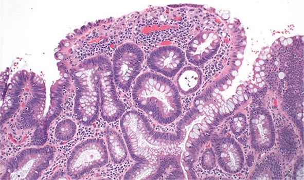
FIGURE 351-13 Medium-power view of low-grade dysplasia in a patient with chronic ulcerative colitis. Low-grade dysplastic crypts are interspersed among regenerating crypts. (Courtesy of Dr. R. Odze, Division of Gastrointestinal Pathology, Department of Pathology, Brigham and Women’s Hospital, Boston, Massachusetts; with permission.)
The risk of neoplasia in chronic UC increases with duration and extent of disease. From one large meta-analysis, the risk of cancer in patients with UC is estimated at 2% after 10 years, 8% after 20 years, and 18% after 30 years of disease. Data from a 30-year surveillance program in the United Kingdom calculated the risk of colorectal cancer to be 7.7% at 20 years and 15.8% at 30 years of disease. The rates of colon cancer are higher than in the general population, and colonoscopic surveillance is the standard of care.
Annual or biennial colonoscopy with multiple biopsies is recommended for patients with >8–10 years of extensive colitis (greater than one-third of the colon involved) or 12–15 years of proctosigmoiditis (less than one-third but more than just the rectum) and has been widely used to screen and survey for subsequent dysplasia and carcinoma. Risk factors for cancer in UC include long-duration disease, extensive disease, family history of colon cancer, PSC, a colon stricture, and the presence of postinflammatory pseudopolyps on colonoscopy.
CROHN’S DISEASE
Risk factors for developing cancer in Crohn’s colitis are long-duration and extensive disease, bypassed colon segments, colon strictures, PSC, and family history of colon cancer. The cancer risks in CD and UC are probably equivalent for similar extent and duration of disease. In the CESAME study, a prospective observational cohort of IBD patients in France, the standardized incidence ratios of colorectal cancer were 2.2 for all IBD patients (95% confidence interval [CI], 1.5–3.0; p < .001) and 7.0 for patients with long-standing extensive colitis (both Crohn’s and UC) (95% CI, 4.4–10.5; p < .001). Thus, the same endoscopic surveillance strategy used for UC is recommended for patients with chronic Crohn’s colitis. A pediatric colonoscope can be used to pass narrow strictures in CD patients, but surgery should be considered in symptomatic patients with impassable strictures.
MANAGEMENT OF DYSPLASIA AND CANCER
Dysplasia can be flat or polypoid. If flat high-grade dysplasia is encountered on colonoscopic surveillance, the usual treatment is colectomy for UC and either colectomy or segmental resection for CD. If flat low-grade dysplasia is found (Fig. 351-13), most investigators recommend immediate colectomy. Adenomas may occur coincidently in UC and CD patients with chronic colitis and can be removed endoscopically provided that biopsies of the surrounding mucosa are free of dysplasia. High-definition and high-magnification colonoscopes and dye sprays have increased the rate of dysplasia detection.
IBD patients are also at greater risk for other malignancies. Patients with CD may have an increased risk of non-Hodgkin’s lymphoma, leukemia, and myelodysplastic syndromes. Severe, chronic, complicated perianal disease in CD patients may be associated with an increased risk of cancer in the lower rectum and anal canal (squamous cell cancers). Although the absolute risk of small-bowel adenocarcinoma in CD is low (2.2% at 25 years in one study), patients with long-standing, extensive, small-bowel disease should consider screening.
352 | Irritable Bowel Syndrome |
Irritable bowel syndrome (IBS) is a functional bowel disorder characterized by abdominal pain or discomfort and altered bowel habits in the absence of detectable structural abnormalities. No clear diagnostic markers exist for IBS; thus the diagnosis of the disorder is based on clinical presentation. In 2006, the Rome II criteria for the diagnosis of IBS were revised (Table 352-1). Throughout the world, about 10–20% of adults and adolescents have symptoms consistent with IBS, and most studies show a female predominance. IBS symptoms tend to come and go over time and often overlap with other functional disorders such as fibromyalgia, headache, backache, and genitourinary symptoms. Severity of symptoms varies and can significantly impair quality of life, resulting in high health care costs. Advances in basic, mechanistic, and clinical investigations have improved our understanding of this disorder and its physiologic and psychosocial determinants. Altered gastrointestinal (GI) motility, visceral hyperalgesia, disturbance of brain-gut interaction, abnormal central processing, autonomic and hormonal events, genetic and environmental factors, and psychosocial disturbances are variably involved, depending on the individual. This progress may result in improved methods of treatment.
DIAGNOSTIC CRITERIA FOR IRRITABLE BOWEL SYNDROMEa |
aCriteria fulfilled for the last 3 months with symptom onset at least 6 months prior to diagnosis. bDiscomfort means an uncomfortable sensation not described as pain. In pathophysiology research and clinical trials, a pain/discomfort frequency of at least 2 days a week during screening evaluation is required for subject eligibility.
Source: Adapted from GF Longstreth et al: Gastroenterology 130:1480, 2006.
CLINICAL FEATURES
IBS is a disorder that affects all ages, although most patients have their first symptoms before age 45. Older individuals have a lower reporting frequency. Women are diagnosed with IBS two to three times as often as men and make up 80% of the population with severe IBS. As indicated in Table 352-1, pain or abdominal discomfort is a key symptom for the diagnosis of IBS. These symptoms should be improved with defecation and/or have their onset associated with a change in frequency or form of stool. Painless diarrhea or constipation does not fulfill the diagnostic criteria to be classified as IBS. Supportive symptoms that are not part of the diagnostic criteria include defecation straining, urgency or a feeling of incomplete bowel movement, passing mucus, and bloating.
Abdominal Pain According to the current IBS diagnostic criteria, abdominal pain or discomfort is a prerequisite clinical feature of IBS. Abdominal pain in IBS is highly variable in intensity and location. It is frequently episodic and crampy, but it may be superimposed on a background of constant ache. Pain may be mild enough to be ignored or it may interfere with daily activities. Despite this, malnutrition due to inadequate caloric intake is exceedingly rare with IBS. Sleep deprivation is also unusual because abdominal pain is almost uniformly present only during waking hours. However, patients with severe IBS frequently wake repeatedly during the night; thus, nocturnal pain is a poor discriminating factor between organic and functional bowel disease. Pain is often exacerbated by eating or emotional stress and improved by passage of flatus or stools. In addition, female patients with IBS commonly report worsening symptoms during the premenstrual and menstrual phases.
Altered Bowel Habits Alteration in bowel habits is the most consistent clinical feature in IBS. The most common pattern is constipation alternating with diarrhea, usually with one of these symptoms predominating. At first, constipation may be episodic, but eventually it becomes continuous and increasingly intractable to treatment with laxatives. Stools are usually hard with narrowed caliber, possibly reflecting excessive dehydration caused by prolonged colonic retention and spasm. Most patients also experience a sense of incomplete evacuation, thus leading to repeated attempts at defecation in a short time span. Patients whose predominant symptom is constipation may have weeks or months of constipation interrupted with brief periods of diarrhea. In other patients, diarrhea may be the predominant symptom. Diarrhea resulting from IBS usually consists of small volumes of loose stools. Most patients have stool volumes of <200 mL. Nocturnal diarrhea does not occur in IBS. Diarrhea may be aggravated by emotional stress or eating. Stool may be accompanied by passage of large amounts of mucus. Bleeding is not a feature of IBS unless hemorrhoids are present, and malabsorption or weight loss does not occur.
Bowel pattern subtypes are highly unstable. In a patient population with ~33% prevalence rates of IBS-diarrhea predominant (IBS-D), IBS-constipation predominant (IBS-C), and IBS-mixed (IBS-M) forms, 75% of patients change subtypes and 29% switch between IBS-C and IBS-D over 1 year. The heterogeneity and variable natural history of bowel habits in IBS increase the difficulty of conducting pathophysiology studies and clinical trials.
Gas and Flatulence Patients with IBS frequently complain of abdominal distention and increased belching or flatulence, all of which they attribute to increased gas. Although some patients with these symptoms actually may have a larger amount of gas, quantitative measurements reveal that most patients who complain of increased gas generate no more than a normal amount of intestinal gas. Most IBS patients have impaired transit and tolerance of intestinal gas loads. In addition, patients with IBS tend to reflux gas from the distal to the more proximal intestine, which may explain the belching.
Some patients with bloating may also experience visible distention with increase in abdominal girth. Both symptoms are more common among female patients and in those with higher overall Somatic Symptom Checklist scores. IBS patients who experienced bloating alone have been shown to have lower thresholds for pain and desire to defecate compared to those with concomitant distention irrespective of bowel habit. When patients were grouped according to sensory threshold, hyposensitive individuals had distention significantly more than those with hypersensitivity and this was observed more in the constipation subgroup. This suggests that the pathogenesis of bloating and distention may not be the same.
Upper Gastrointestinal Symptoms Between 25 and 50% of patients with IBS complain of dyspepsia, heartburn, nausea, and vomiting. This suggests that other areas of the gut apart from the colon may be involved. Prolonged ambulant recordings of small-bowel motility in patients with IBS show a high incidence of abnormalities in the small bowel during the diurnal (waking) period; nocturnal motor patterns are not different from those of healthy controls. The overlap between dyspepsia and IBS is great. The prevalence of IBS is higher among patients with dyspepsia (31.7%) than among those who reported no symptoms of dyspepsia (7.9%). Conversely, among patients with IBS, 55.6% reported symptoms of dyspepsia. In addition, the functional abdominal symptoms can change over time. Those with predominant dyspepsia or IBS can flux between the two. Although the prevalence of functional gastrointestinal disorders is stable over time, the turnover in symptom status is high. Many episodes of symptom disappearance are due to subjects changing symptoms rather than total symptom resolution. Thus it is conceivable that functional dyspepsia and IBS are two manifestations of a single, more extensive digestive system disorder. Furthermore, IBS symptoms are prevalent in noncardiac chest pain patients, suggesting overlap with other functional gut disorders.
PATHOPHYSIOLOGY
The pathogenesis of IBS is poorly understood, although roles of abnormal gut motor and sensory activity, central neural dysfunction, psychological disturbances, mucosal inflammation, stress, and luminal factors have been proposed.
Gastrointestinal Motor Abnormalities Studies of colonic myoelectrical and motor activity under unstimulated conditions have not shown consistent abnormalities in IBS. In contrast, colonic motor abnormalities are more prominent under stimulated conditions in IBS. IBS patients may exhibit increased rectosigmoid motor activity for up to 3 h after eating. Similarly, inflation of rectal balloons both in IBS-D and IBS-C patients leads to marked and prolonged distention-evoked contractile activity. Recordings from the transverse, descending, and sigmoid colon showed that the motility index and peak amplitude of high-amplitude propagating contractions (HAPCs) in diarrhea-prone IBS patients were greatly increased compared to those in healthy subjects and were associated with rapid colonic transit and accompanied by abdominal pain.
Visceral Hypersensitivity As with studies of motor activity, IBS patients frequently exhibit exaggerated sensory responses to visceral stimulation. The frequency of perceptions of food intolerance is at least twofold more common than in the general population. Postprandial pain has been temporally related to entry of the food bolus into the cecum in 74% of patients. On the other hand, prolonged fasting in IBS patients is often associated with significant improvement in symptoms. Rectal balloon inflation produces nonpainful and painful sensations at lower volumes in IBS patients than in healthy controls without altering rectal tension, suggestive of visceral afferent dysfunction in IBS. Similar studies show gastric and esophageal hypersensitivity in patients with nonulcer dyspepsia and noncardiac chest pain, raising the possibility that these conditions have a similar pathophysiologic basis. Lipids lower the thresholds for the first sensation of gas, discomfort, and pain in IBS patients. Hence, postprandial symptoms in IBS patients may be explained in part by a nutrient-dependent exaggerated sensory component of the gastrocolonic response. In contrast to enhanced gut sensitivity, IBS patients do not exhibit heightened sensitivity elsewhere in the body. Thus, the afferent pathway disturbances in IBS appear to be selective for visceral innervation with sparing of somatic pathways. The mechanisms responsible for visceral hypersensitivity are still under investigation. It has been proposed that these exaggerated responses may be due to (1) increased end-organ sensitivity with recruitment of “silent” nociceptors; (2) spinal hyperexcitability with activation of nitric oxide and possibly other neurotransmitters; (3) endogenous (cortical and brainstem) modulation of caudad nociceptive transmission; and (4) over time, the possible development of long-term hyperalgesia due to development of neuroplasticity, resulting in permanent or semipermanent changes in neural responses to chronic or recurrent visceral stimulation (Table 352-2).
PROPOSED MECHANISMS FOR VISCERAL HYPERSENSITIVITY |
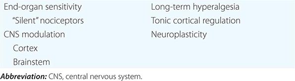
Central Neural Dysregulation The role of central nervous system (CNS) factors in the pathogenesis of IBS is strongly suggested by the clinical association of emotional disorders and stress with symptom exacerbation and the therapeutic response to therapies that act on cerebral cortical sites. Functional brain imaging studies such as magnetic resonance imaging (MRI) have shown that in response to distal colonic stimulation, the mid-cingulate cortex—a brain region concerned with attention processes and response selection—shows greater activation in IBS patients. Modulation of this region is associated with changes in the subjective unpleasantness of pain. In addition, IBS patients also show preferential activation of the prefrontal lobe, which contains a vigilance network within the brain that increases alertness. These may represent a form of cerebral dysfunction leading to the increased perception of visceral pain.
Abnormal Psychological Features Abnormal psychiatric features are recorded in up to 80% of IBS patients, especially in referral centers; however, no single psychiatric diagnosis predominates. Most of these patients demonstrated exaggerated symptoms in response to visceral distention, and this abnormality persists even after exclusion of psychological factors.
Psychological factors influence pain thresholds in IBS patients, as stress alters sensory thresholds. An association between prior sexual or physical abuse and development of IBS has been reported. Abuse is associated with greater pain reporting, psychological distress, and poor health outcome. Brain functional MRI studies show greater activation of the posterior and middle dorsal cingulate cortex, which is implicated in affect processing in IBS patients with a past history of sexual abuse.
Thus, patients with IBS frequently demonstrate increased motor reactivity of the colon and small bowel to a variety of stimuli and altered visceral sensation associated with lowered sensation thresholds. These may result from CNS–enteric nervous system dysregulation (Fig. 352-1).
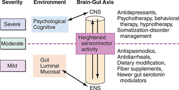
FIGURE 352-1 Therapeutic targets for irritable bowel syndrome. Patients with mild to moderate symptoms usually have intermittent symptoms that correlate with altered gut physiology. Treatments include gut-acting pharmacologic agents such as antispasmodics, antidiarrheals, fiber supplements, and gut serotonin modulators. Patients who have severe symptoms usually have constant pain and psychosocial difficulties. This group of patients is best managed with antidepressants and other psychosocial treatments. CNS, central nervous system; ENS, enteric nervous system.
Postinfectious IBS IBS may be induced by GI infection. In an investigation of 544 patients with confirmed bacterial gastroenteritis, one-quarter developed IBS subsequently. Conversely, about a third of IBS patients experienced an acute “gastroenteritis-like” illness at the onset of their chronic IBS symptomatology. This group of “postinfective” IBS occurs more commonly in females and affects younger rather than older patients. Risk factors for developing postinfectious IBS include, in order of importance, prolonged duration of initial illness, toxicity of infecting bacterial strain, smoking, mucosal markers of inflammation, female gender, depression, hypochondriasis, and adverse life events in the preceding 3 months. Age older than 60 years might protect against postinfectious IBS, whereas treatment with antibiotics has been associated with increased risk. The microbes involved in the initial infection are Campylobacter, Salmonella, and Shigella. Those patients with Campylobacter infection who are toxin-positive are more likely to develop postinfective IBS. Increased rectal mucosal enteroendocrine cells, T lymphocytes, and increased gut permeability are acute changes following Campylobacter enteritis that could persist for more than a year and may contribute to postinfective IBS.
Immune Activation and Mucosal Inflammation Some patients with IBS display persistent signs of low-grade mucosal inflammation with activated lymphocytes, mast cells, and enhanced expression of proinflammatory cytokines. These abnormalities may contribute to abnormal epithelial secretion and visceral hypersensitivity. There is increasing evidence that some members of the superfamily of transient receptor potential (TRP) cation channels such as TRPV1 (vanilloid) channels are central to the initiation and persistence of visceral hypersensitivity. Mucosal inflammation can lead to increased expression of TRPV1 in the enteric nervous system. Enhanced expression of TRPV1 channels in the sensory neurons of the gut has been observed in IBS, and such expression appears to correlate with visceral hypersensitivity and abdominal pain. Interestingly, clinical studies have also shown increased intestinal permeability in patients with IBS-D. Psychological stress and anxiety can increase the release of proinflammatory cytokine, and this in turn may alter intestinal permeability. This provides a functional link between psychological stress, immune activation, and symptom generation in patients with IBS.
Altered Gut Flora A high prevalence of small intestinal bacterial overgrowth in IBS patients has been noted based on positive lactulose hydrogen breath test. This finding, however, has been challenged by a number of other studies that found no increased incidence of bacterial overgrowth based on jejunal aspirate culture. Abnormal H2 breath test can occur because of small-bowel rapid transit and may lead to erroneous interpretation. Hence, the role of testing for small intestinal bacterial overgrowth in IBS patients remains unclear.
Studies using culture-independent approaches such as 16S rRNA gene-based analysis found significant differences between the molecular profile of the fecal microbiota of IBS patients and that of healthy subjects. IBS patients had decreased proportions of the genera Bifidobacterium and Lactobacillus and increased ratios of Firmicutes:Bacteroidetes. It has been speculated that these changes may be related to stress and diet. A temporary reduction in lactobacilli has been reported in animal models of early-life stress. On the other hand, Firmicutes is the dominant phylum in adults consuming a diet high in animal fat and protein. However, it is still unclear whether such changes in fecal microbiota are causal, consequential, or merely the result of constipation and diarrhea. In addition, the stability of the change in the microbiota needs to be determined.
Abnormal Serotonin Pathways The serotonin (5-HT)-containing enterochromaffin cells in the colon are increased in a subset of IBS-D patients compared to healthy individuals or patients with ulcerative colitis. Furthermore, postprandial plasma 5-HT levels were significantly higher in this group of patients compared to healthy controls. Because serotonin plays an important role in the regulation of GI motility and visceral perception, the increased release of serotonin may contribute to the postprandial symptoms of these patients and provides a rationale for the use of serotonin antagonists in the treatment of this disorder.


