May be difficult to identify, especially if desmoplastic type or heavily inflamed
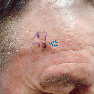
The marked area
 , prior to biopsy, shows a papular growth in an ill-defined pigmented patch. This raises concern for a microinvasive melanoma. (Courtesy J. Finch, MD.)
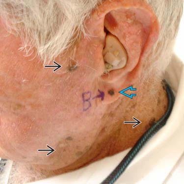
The marked lesion is concerning for melanoma
 . This lesion stands out among the many seborrheic keratoses
  on the left face and neck. (Courtesy J. Finch, MD.)
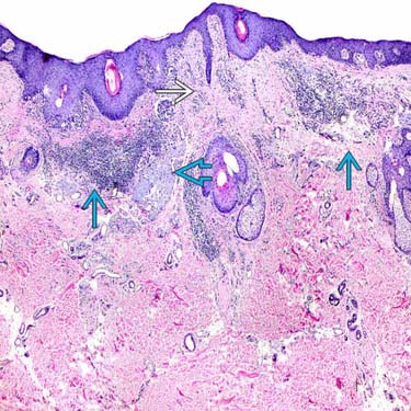
This broad, atypical melanocytic proliferation displays fibrosis
 and patchy, inflammatory infiltrate
  , consistent with inflammatory regression. Even the solar elastosis
  is interrupted in the area of fibrosis.
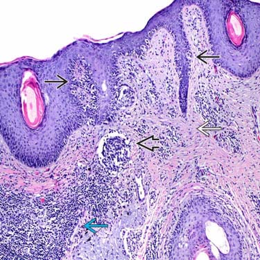
Above the lentiginous atypical, melanocytic proliferation
 , there is a prominent invasive nest
  in the papillary dermis. Note the fibrosis (interrupted solar elastosis)
  and inflammation
  .
.


MICROSCOPIC
Histologic Features
• Severely atypical compound melanocytic proliferation with junctional lentiginous component (typical of lentigo maligna)


Stay updated, free articles. Join our Telegram channel
Full access? Get Clinical Tree
