• Irritated/traumatized nevi and nevi of special sites
• Reed (pigmented spindle cell) nevus
Diagnostic Checklist
• Single atypical melanocytes as well as irregular nesting and areas of limited pagetoid scatter
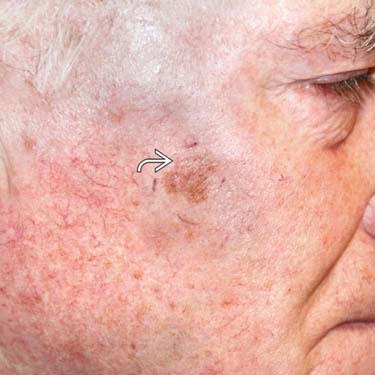
Clinical Photograph of MIS, NOS This lightly pigmented patch on chronically sun-exposed skin has irregular borders. It appears to be ill-defined, and a biopsy is needed to rule in melanoma. The biopsy proved a melanoma in situ (MIS). The lesion is evenly pigmented; however, there are less pigmented foci , suggestive of early regression.
Irregular Patch on Sun-Exposed Skin There is a growing pigmented patch (circled) in a background of sun damage, lentigines, and actinic keratoses. The ill-defined patch should raise concern and be biopsied. (Courtesy J. Finch, MD.)
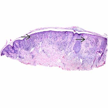
MIS Showing Hypercellular Junctional Growth Under scanning magnification, there is an overgrowth of intraepidermal melanocytes . This configuration should raise concern for MIS, even at scanning magnification.
MIS Showing Upward Scatter of Melanocytes There is migration of melanocytes throughout all the epidermal layers . A patchy lymphocytic infiltrate , telangiectasia , and solar elastosis are noted. Note how the melanocytes outnumber the keratinocytes.
TERMINOLOGY
Abbreviations
• Melanoma in situ, not otherwise specified (MIS, NOS)
Synonyms
• Melanoma in situ, unspecified type
Definitions
• Cases that cannot be clearly categorized as either lentigo maligna, acral lentiginous, or superficial spreading type of melanoma in situ
CLINICAL ISSUES
Epidemiology
• Incidence
Uncommon tumors
• Age
Typically occur in elderly patients
Site
• Usually located on sun-exposed areas, especially cheeks, upper trunk, and arms
Presentation
• Borders of lesions are irregular, asymmetric, and often notched
• Lesions are dark, broad macules ~ 0.8 cm in diameter
• Mottled pigmentation
Treatment
• Surgical approaches
Complete excision with 5.0-mm clear margins
Mohs surgery may also be effective, but is controversial
Prognosis
• Excellent: Little recurrence potential with adequate surgical margins
MICROSCOPIC
Histologic Features
• Melanocytic proliferation typically characterized by both lentiginous junctional spread (similar to lentigo maligna) and irregular nesting and pagetoid spread (similar to superficial spreading type of MIS)
 , suggestive of early regression.
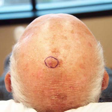
 . This configuration should raise concern for MIS, even at scanning magnification.
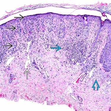
 . A patchy lymphocytic infiltrate
  , telangiectasia
  , and solar elastosis
  are noted. Note how the melanocytes outnumber the keratinocytes.


