Mammary Carcinoma with Osteoclast-like Giant Cells
FREDERICK C. KOERNER
Carcinomas containing osteoclast-like multinucleated giant cells arise in many organs, including the breast, lung, pancreas, small intestine, and thyroid gland. Similar giant cells have also been found in noncarcinomatous tumors such as uterine leiomyosarcoma1 and intestinal carcinoid.2 In 1979, Agnantis and Rosen3 first documented the presence of osteoclast-like giant cells in mammary carcinomas, and since then approximately 200 examples of this type of mammary carcinoma have been reported.
CLINICAL PRESENTATION
Despite the unusual histologic properties of these tumors, the clinical features are similar to those of breast carcinoma generally. Patients’ age ranged from 28 to 88 years, and the average age at diagnosis was approximately 50 years.3,4,5,6 Typically, the patient presented with a palpable tumor in the upper outer quadrant, but the lesion has been found in all quadrants. Multifocal lesions were described clinically in two cases.7,8 Bilateral primary carcinomas with osteoclastlike giant cells are exceedingly rare.9 On mammography and ultrasonography, the well-circumscribed margin of most tumors may suggest a benign lesion such as a cyst or a fibroadenoma (FA).5,10 Masses with irregular margins and inhomogeneous internal echoes have been reported.8 Magnetic resonance imaging (MRI) of one carcinoma exhibited “rich vascularity, especially in the periphery.”11
GROSS PATHOLOGY
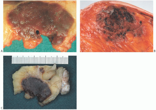
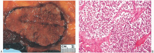
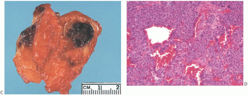
The tumors are usually well defined, fleshy, and firm. Reported diameters range from 0.5 to 10 cm, with most carcinomas measuring 3 cm or less. The macroscopic appearance of most tumors is quite striking: when bisected, the dark brown or red-brown tumor tends to bulge slightly above the surrounding parenchyma from which it may be separated by a rounded, discrete margin (Fig. 23.1). Tumors with illdefined margins and multinodules have been described.7,12 The deep mahogany color may suggest heavily pigmented, metastatic malignant melanoma, but the color of carcinomas with osteoclast-like giant cells tends to be brown rather than black. Tumors with relatively few osteoclast-like giant cells or with little hemorrhage may appear tan or white. These unusual features are not specific for this neoplasm because some solid papillary or nonmedullary circumscribed carcinomas that lack giant cells microscopically are grossly indistinguishable from carcinomas with osteoclast-like giant cells (Fig. 23.2).
MICROSCOPIC PATHOLOGY
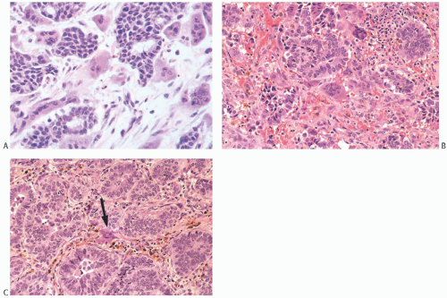
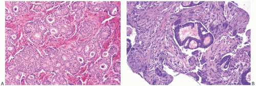
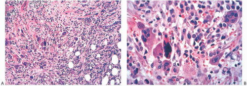
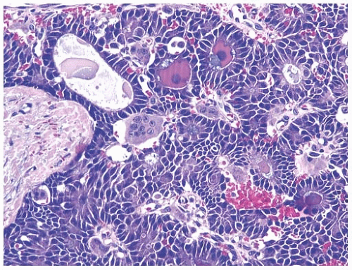
Most of these lesions are moderately or poorly differentiated invasive duct carcinomas (Fig. 23.3). A cribriform growth pattern is present relatively more often than occurs among duct carcinomas generally (Fig. 23.4). Uncommon examples of well-differentiated or tubular4,12 (Fig. 23.5), lobular3,9,10,13 (Fig. 23.6), squamous,14 papillary3 (Fig. 23.7), apocrine (Fig. 23.8), mucinous6 (Fig. 23.9), metaplastic,12 and neuroendocrine15 carcinomas with osteoclast-like giant cells have been described. Rarely, the carcinoma has a glandular pattern reminiscent of that of infiltrating colonic carcinoma (Fig. 23.10). Osteoclast-like giant cells can be encountered in anaplastic carcinomas that are probably variants of metaplastic carcinoma (Fig. 23.11). When present, the ductal carcinoma in situ (DCIS) has the appearance of one of the conventional variants, usually cribriform, solid, or papillary. Osteoclast-like giant cells are not always present in the associated DCIS (Fig. 23.11). It is very uncommon to find osteoclast-like giant cells in DCIS in the absence of an invasive lesion (Fig. 23.12).16
The osteoclast-like giant cells range from 20 to 180 µm in diameter.17 They contain abundant cytoplasm and many evenly distributed and usually centrally located oval nuclei, some of which contain small nucleoli. The giant cells tend to cluster close to the edges of carcinomatous glands or in intervening stroma, and they may be found in the glandular lumens (Figs. 23.7, 23.9, and 23.10). The stroma typically contains mononuclear histiocytes whose cytological features resemble those of the multinuclear giant cells.
The presence of extravasated erythrocytes and hemosiderin, which one finds in the vascular stroma in most cases, reflects recent and older episodes of hemorrhage (Figs. 23.3, 23.5, and 23.6). Erythrophagocytosis by the giant cells is uncommon, and they contain little hemosiderin detectable by light microscopy. Fibroblastic reaction, collagenization, angiogenesis, and lymphocytic infiltration are variably present in the stroma (Figs. 23.3, 23.4, and 23.13).
Osteoclast-like giant cells are found in examples of metaplastic carcinoma that contain areas of osseous and cartilaginous differentiation18,19; conversely, inconspicuous and infrequent metaplastic foci with spindle cells and squamous or osseous features have been described in tumors that otherwise were typical examples of mammary carcinoma with osteoclast-like giant cells.3,12 Mammary carcinoma with osteoclast-like giant cells may be a variant of metaplastic mammary carcinoma, but it seems appropriate to separate these tumors until investigators have better defined the clinicopathologic characteristics of these two types of mammary carcinoma.
 FIG. 23.5. Mammary carcinoma with osteoclast-like giant cells, well differentiated. A,B: The stroma in this tumor contains many lymphocytes and red blood cells. |
Differential Diagnosis
The differential diagnosis includes certain noncarcinomatous lesions.20 Megakaryocytes in myeloid metaplasia in the breast might be mistaken for osteoclast-like giant cells, but these lesions have abundant myeloid elements in various stages of maturation, a feature not found in carcinomas with osteoclast-like giant cells.21 Granulomatous foci in inflammatory conditions such as sarcoidosis or coexistent with carcinoma also contain giant cells that differ histologically from those with osteoclast-like features22,23 (Fig. 23.14). Because mammary carcinoma with osteoclast-like giant cells does not have a granulomatous pattern, the distinction from sarcoid or tuberculoid reactions can be made without difficulty in histologic sections (see Chapter 4). The osteoclast-like giant cells found in mammary carcinoma do not resemble the multinucleated stromal giant cells that are an incidental finding in breast tissue from patients with benign conditions or carcinoma (Fig. 23.15). Multinucleated stromal giant cells may also be found in the stroma of fibroepithelial tumors (see Chapter 8). They lack the relatively abundant cytoplasm seen in osteoclast-like giant cells.
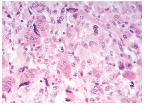 FIG. 23.8. Mammary carcinoma with osteoclast-like giant cells, apocrine. The giant cells mingle with apocrine carcinoma cells, which have prominent nucleoli. |
Stay updated, free articles. Join our Telegram channel

Full access? Get Clinical Tree


