Malignant Mesothelioma
Cyril Fisher, MD, DSc, FRCPath
Key Facts
Terminology
Malignant tumor of mesothelial cells arising in pleura, peritoneum, or pericardium
Etiology/Pathogenesis
Asbestos exposure
Amphibole: Crocidolite, amosite
Thin long fibers most oncogenic
Clinical Issues
5-10x more common in males
Invades underlying organs and adjacent structures
Metastasizes
Microscopic Pathology
Sheet-like, tubular, papillary, microglandular, or mixed patterns
Fibrosarcoma-like fascicles
Top Differential Diagnoses
Reactive mesothelial proliferation
No atypia, necrosis, invasion
Desmin(+), EMA(-)
Adenocarcinoma of lung
BER-EP4, MOC-31, CEA, CD15, TTF-1 (some) typically positive
Calretinin, thrombomodulin, CK5/6, WT1 typically negative
Synovial sarcoma
Solitary fibrous tumor
No pattern
CD34(+)
Cytokeratin, EMA typically negative
Angiosarcoma
CD34, CD31, FLI-1 positive
TERMINOLOGY
Abbreviations
Malignant mesothelioma (MM)
Synonyms
Diffuse mesothelioma
Definitions
Malignant tumor of mesothelial cells arising in pleura, peritoneum, or pericardium
ETIOLOGY/PATHOGENESIS
Environmental Exposure
Asbestos
Occupational exposure
Mining, construction, vehicle maintenance, shipbuilding
Risk relates to intensity and duration of exposure
Amphibole fiber types most carcinogenic
Persist in lung with or without tissue reaction
Crocidolite and amosite have highest risk
Longer, thinner fibers more oncogenic
Latent period: 15-40 years
Occasionally other fiber types implicated, e.g., erionite
Infectious Agents
SV40 DNA oncogenic virus
Viral sequences found in some mesotheliomas
Contaminated polio vaccine unproven association
Radiation
Therapeutic
Contrast media, e.g., thorium dioxide
Chronic Inflammation
Rarely after prolonged fibrosing inflammation, e.g., empyema
CLINICAL ISSUES
Epidemiology
Incidence
Geographic variation
In USA, ˜ 20 cases per million of population in males, now peaked and declining
In UK, Australia, incidence higher and increasing
In Europe, incidence beginning to plateau
Age
Most > 60 years
Occasional cases at any age
Gender
5-10x more common in males
Only 20% of cases in females are asbestos-related
Presentation
Dyspnea, chest pain, cough, weight loss
Abdominal pain, distension, vague mass
Natural History
Multiple small lesions fuse to form diffuse sheet
Invades underlying organs and adjacent structures
Lung, mediastinum, through diaphragm, chest wall
Viscera, abdominal wall
Metastasizes
To lung, lymph nodes
Rarely to liver, bone, brain, kidney, adrenal
Treatment
Options, risks, complications
Multimodal therapy more effective than single
Surgical approaches
Extrapleural pneumonectomy
Decortication
Adjuvant therapy
Chemotherapy and radiotherapy can prolong survival
More often palliative
Prognosis
Poor
Median survival: 7 months; mortality: 100%
Desmoplastic sarcomatoid variant is most aggressive
IMAGE FINDINGS
General Features
Pleural effusion
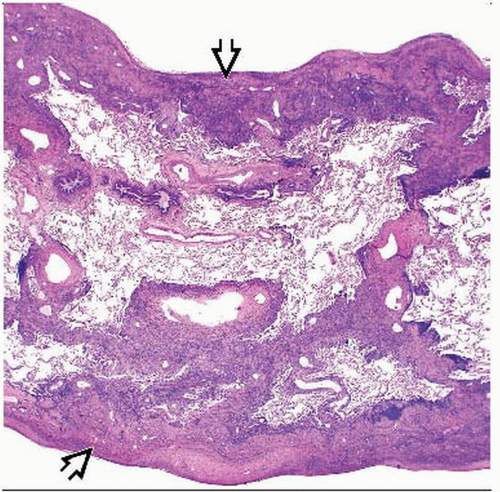
Diffuse pleural thickening extending into fissures
Peritoneal thickening
Mass
MACROSCOPIC FEATURES
General Features
Multiple small nodules grow and coalesce
Firm diffuse white tumor o Encases lung
Locules contain fluid
Invades adjacent structures
Lung, mediastinum, diaphragm
Chest wall, abdominal wall
MICROSCOPIC PATHOLOGY
Histologic Features
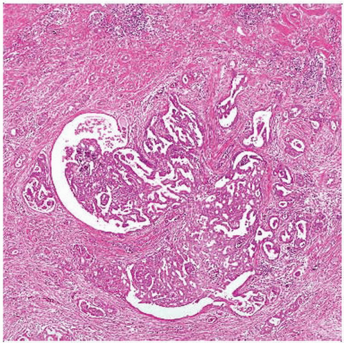
Epithelioid malignant mesothelioma
Polygonal, cuboidal, or flattened discohesive cells
Cytoplasm eosinophilic, rarely clear
Mild nuclear pleomorphism, variable mitoses
Sheet-like, tubular, papillary, microglandular, or mixed patterns
Psammoma bodies
Variable stromal collagen, inflammation, rare myxoid change
Asbestos bodies in adjacent tissues
Sarcomatoid malignant mesothelioma
Fibrosarcoma-like fascicles
Variably pleomorphic spindle cells
Giant cells
Fibrosis, hyalinization (desmoplastic variant)
Necrosis
Heterologous osteosarcoma, chondrosarcoma, rhabdomyosarcoma
Biphasic malignant mesothelioma
Mixed epithelioid and sarcomatoid morphology
Other histological variants of malignant mesothelioma
Small cell
Clear cell
Deciduoid
Lymphohistiocytoid
Pleomorphic
Predominant Pattern/Injury Type
Diffuse
Predominant Cell/Compartment Type


