Malignant Cellular Blue Nevus (Melanoma Arising in Cellular Blue Nevus)
Symmetric and well circumscribed
Often shows dumbbell-like appearance at low power
Cells lack prominent cytologic atypia, mitotic activity, and necrosis
• Atypical cellular blue nevus
Shows overlapping features with cellular blue nevus, but with increased cytologic atypia
May have some mitoses, but not numerous or atypical
• Metastatic melanoma
Usually mostly epithelioid-appearing melanocytes, fewer spindled cells, and no dendritic cells
No associated benign blue nevus component
Malignant Cellular Blue Nevus With Necrosis This was a very large and deep cellular lesion in an older adult patient. Note the central focus of necrosis surrounded by cellular nodules of hyperchromatic-appearing cells.
Densely Cellular Fascicles of Spindle Cells in Malignant Cellular Blue Nevus This malignant blue nevus shows densely cellular areas of atypical spindled melanocytes forming fascicular (sarcoma-like) arrangements.
High Magnification of Heavily Pigmented Malignant Cellular Blue Nevus High magnification of a heavily pigmented malignant cellular blue nevus (MCBN) demonstrates significant nuclear atypia with prominent nucleoli and a mitotic figure in this case.
Ki-67 Immunostaining in Malignant Cellular Blue Nevus Ki-67 immunostaining shows increased nuclear positivity in this malignant cellular blue nevus, with scattered enlarged, atypical-appearing nuclei staining .
TERMINOLOGY
Abbreviations
• Malignant cellular blue nevus (MCBN)
Synonyms
• Melanoma arising in cellular blue nevus
• Melanoma mimicking cellular blue nevus
Definitions
• Malignant dermal-based melanocytic neoplasm with associated cellular blue nevus or features simulating cellular blue nevus
ETIOLOGY/PATHOGENESIS
Environmental Exposure
• May be related to solar damage
Genetics
• Multiple chromosomal abnormalities reported in some cases
Gains and losses of entire chromosomal arms are most common aberrations
• Mutations in GNAQ and GNA11 have been reported
• Loss of nuclear BAP1 expression also recently identified
• 1 case reported to show mutation in hOGG-1 DNA repair enzyme
CLINICAL ISSUES
Epidemiology
• Incidence
Very rare tumor
• Age
Usually occurs in adults
• Sex
More common in males
Presentation
• Dermal mass/nodular lesion
May appear blue, bluish-gray, or bluish-black
Treatment
• Surgical approaches
Complete and wider surgical excision is necessary for local removal
Sentinel lymph node biopsy may be used for staging purposes but is of doubtful therapeutic utility
Prognosis
• Controversial
Originally thought to be very aggressive, but recent reports may indicate lower malignant potential
Only gold members can continue reading. Log In or Register to continue
Apr 24, 2017 | Posted by admin in PATHOLOGY & LABORATORY MEDICINE | Comments Off on Malignant Cellular Blue Nevus (Melanoma Arising in Cellular Blue Nevus)


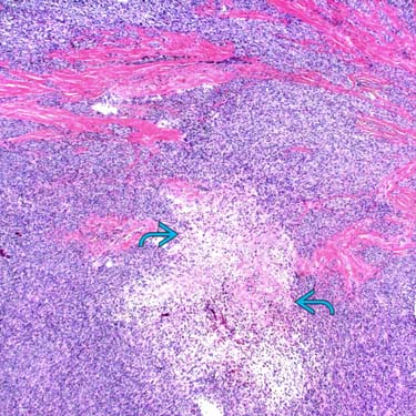
 surrounded by cellular nodules of hyperchromatic-appearing cells.
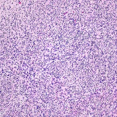
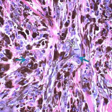
 and a mitotic figure
  in this case.
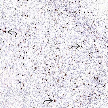
 .
.


