Magnesium1


Magnesium1


Robert K. Rude


Deceased.


Magnesium (Mg) plays an essential role in a wide range of fundamental biologic reactions. Hence, it is not surprising that Mg deficiency may lead to serious clinical symptoms. Kruse et al (1) made the first systematic observations of Mg deficiency in rats and dogs in the early 1930s. The first description of clinical depletion in humans, published in 1934, involved a small number of patients with various underlying diseases (2). In the early 1950s, Flink (3) initiated studies documenting depletion of this ion in patients with alcoholism and in patients receiving Mg-free intravenous solutions. Although the diets ordinarily consumed by healthy Americans contain less Mg than the recommended dietary allowance (RDA) (4), they do not appear to lead to symptomatic Mg depletion. Some clinical disorders, however, as discussed in this chapter, have been associated with a low-Mg diet.


BIOCHEMISTRY AND PHYSIOLOGY

Mg is widely distributed in nature, and it is the eighth most abundant element on earth and the second most abundant cation in sea water (5, 6). Mg is the fourth most abundant cation in the body and the second most prevalent intracellular cation (5, 6). Because of its positive charge, Mg binds to negatively charged molecules. Most intracellular Mg binds to ribosomes, membranes, and other macromolecules in the cytosol and nucleus.


Enzyme Interactions

Mg is involved in more than 300 essential metabolic reactions (7). The Mg ion (Mg2+) forms complexes with a variety of organic molecules. Mg2+ is essential for many enzymatic reactions and has two general interactions: (a) Mg2+ binds to the substrate, thereby forming a complex with which the enzyme interacts, as in the reaction of kinases with Mg adenosine triphosphate (MgATP); and (b) Mg2+ binds directly to the enzyme and alters its structure or serves a catalytic role (e.g., exonuclease, topoisomerase, and RNA and DNA polymerases) (6, 8, 9). Overall, the predominant action of Mg is related to ATP utilization. ATP has a strategic position in “freeenergy” currency for virtually all cellular processes, by providing high-energy phosphate. It exists in all cells,
primarily as MgATP2-. Mg therefore is essential for the function of the glycolytic cycle, citric acid cycle, protein kinases, RNA and DNA polymerases, lipid metabolism, and amino acid activation, as well as playing a critical role in the cyclic adenosine monophosphate (cAMP) and phospholipase C second-messenger systems (5, 6, 10, 11, 12).


Structural Modification of Nucleic Acids and Membrane

Another important role of Mg is its ability to form complexes with nucleic acids. The negatively charged ribose phosphate structure of nucleic acids has a high affinity for Mg2+; the resulting stabilization of numerous ribonucleotides and deoxyribonucleotides induces important physicochemical changes that affect DNA maintenance, duplication, and transcription. (6, 7, 8, 9, 13). In addition, the binding of hydrated Mg2+ by transfer RNA (tRNA) and modified tRNA and its DNA analogs results in structures that cannot be duplicated by the binding of other metals (6, 7, 8, 9, 13).

Mg, calcium (Ca2+), and some other cations react with hydrophilic polyanionic carboxylates and phosphates of the various membrane components to stabilize the membrane and thereby affect fluidity and permeability. This process influences ion channels, transporters, and signal transducers (6).


Ion Channels

Ion channels constitute a class of proteins across the cell membrane, which allow passage of ions into or out of cells when the channels are open. Ion channels are classified according to the type of ion they allow to pass, such as sodium (Na+), potassium (K+), or Ca2+ (14). Mg2+ plays an important role in the function of certain ion channels. A deficit of Mg results in cellular potassium (K) depletion (14). Mg2+ is necessary for the active transport of K+ out of cells by Na+/K+-ATPase (15). Another mechanism for K+ loss is an increased efflux of K+ from cells via other Mg2+-sensitive K+ channels, as has been seen in skeletal and heart muscle (16, 17). Therefore, deficiency in Mg2+ leads to reduced intracellular K+. The arrhythmogenic effect of Mg deficiency, as discussed later, may be related to its effect on intracellular K+.

Mg has been called nature’s physiologic Ca channel blocker (14). During Mg depletion, intracellular Ca2+ rises. This may be the result of both an increase from extracellular Ca2+ and release from intracellular Ca2+ stores. Mg2+ has been demonstrated to decrease the inward Ca2+ flux through slow Ca channels (15). In addition, Mg2+ decreases the transport of Ca2+ out of the sarcoplasmic reticulum into the cell cytosol. Inositol triphosphate (IP3) has an inverse ability to release Ca2+ from intracellular stores in response to changes in Mg2+ concentrations, and this also contributes to a rise in intracellular Ca2+ during a fall in Mg2+ (12).


BODY COMPOSITION AND HOMEOSTASIS


Composition

The distribution of Mg in various body compartments of apparently healthy adult individuals is summarized in Table 9.1. Approximately 60% of Mg is in the skeleton— two thirds is within the hydration shell, and one third is on the crystal surface (18). This may serve as a reservoir for maintaining extracellular and intracellular Mg. Only 1% of Mg is in the extracellular fluid; the rest is intracellular (19).


TABLE 9.1 DISTRIBUTION AND CONCENTRATIONS OF MAGNESIUM (Mg) IN A HEALTHY ADULT (TOTAL BODY: 833-1170 mmola, OR 20-28 g)


SITE


PERCENTAGE OF TOTAL BODY Mg (%)


CONCENTRATION/CONTENT


Bone


53


0.5% of bone ash


Muscle


27


9 mmol/kg wet weight


Soft tissue


19


9 mmol/kg wet weight


Adipose tissue


0.012


0.8 mmol/kg wet weightb


Erythrocytes


0.5


1.65-2.73 mmol/Lc


Serum


0.3


0.88 ± 0.06 mml/Ld


Percentage of total


Free


65


0.56 ± 0.05 m/mol/Le


Complexed


8


Bound


27


Mononuclear


2.91 ± 0.6 fmol/cellg


Blood cellsf


2.79 ± 0.6 fmol/cellh


3.00 ± 0.4 fmol/celli


Platelets


2.26 ± 0.29 mmol/Lj


[Mg2+]ik


0.5-1.0 mmol/L


Cerebrospinal fluid


1.25 mmol/L


Free 55%


Complexed 45%


Secretions


Saliva, gastric, bile


0.3-0.7 mmol/L


Sweat


0.3 mmol/L (38°C)l


0.09 mmol/hm


a1 mmol = 2 mEq = 24.3 mg.

b From Snyder WS. Report of the Task Group on Reference Man. Elmsford, NY: Pergamon Press, 1975:306.

c Magnesium falls slowly with aging.

d Similar at various ages.

e From Huijgen HJ, Van Ingen HE, Kok WT et al. Clin Biochem 1996;29:261-6.

f Monocytes and lymphocytes in venous blood.

g From Elin RJ, Hosseini JM. Clin Chem 1985;31:377-80. 1 fmol = 24.3 fg.

h From Reinhart RA, Marx JJ Jr, Haas RG et al. Clin Chim Acta 1987;167:187-95.

i From Yang XY, Hosseini JM, Ruddel ME, Elin RJ. J Am Coll Nutr 1990;9:328.

j From Niemala JE, Snader BM, Elin RJ. Clin Chem 1996;42:744-8.

k Intracellular free magnesium concentration.

l From Consolazio CF, Matoush LO, Nelson RA et al. J Nutr 1963;79:407.

m From Wenk C, Kuhnt M, Kunz P et al. Methodological studies of the estimation of loss of sodium, potassium, calcium and magnesium through the skin during a 10 km run [in German]. Z Ernahrungswiss 1993;32:301-7.


Cellular Homeostasis

Mg is compartmentalized within the cell, and most of it is bound to proteins and negatively charged molecules. Significant amounts of Mg are found in the nucleus, mitochondria, the endoplasmic and sarcoplasmic reticulum, and the cytoplasm (5, 6, 20). Total cell Mg concentration has been reported to range between 5 and 20 mM (15). From 90% to 95% of cytosolic Mg is bound to ligands such as ATP, adenosine diphosphate (ADP), citrate, proteins, and nucleic acids. The remainder is free Mg2+, constituting 1% to 5% of the total cellular Mg (15, 21).

The concentration of free ionized Mg2+ in the cytoplasm of mammalian cells has ranged from 0.5 to 1.0 mM, similar to circulating ionized Mg2+ (6, 15). The Mg2+ concentration in the cell cytoplasm is maintained relatively constant even when the Mg2+ concentration in the extracellular fluid is experimentally varied to either high or low nonphysiologic concentrations (22). The relative constancy of the Mg2+ in the intracellular milieu is attributed to the limited permeability of the plasma membrane to Mg and to the operation of specific Mg transport proteins, which regulate the rates at which Mg is taken up or extruded from cells (5, 6, 15). Maintenance of a normal intracellular concentration of Mg2+ requires that Mg be actively transported out of the cell (15). Mg transport into or out of cells appears to require the presence of carriermediated transport systems. The efflux of Mg from the cell appears to be coupled to Na transport and requires extrusion of Na by Na+/K+-ATPase (15). Evidence also indicates a Na-independent efflux of Mg (7, 15). Mg influx appears to be linked to Na transport, but by a different mechanism than efflux (15, 23). At least seven transmembrane Mg2+ channels have been cloned (24). These include NIPA2 (25) and MagT1 and TUSC3 (26). Studies of human hereditary diseases (see later) have identified paracellin-1 (claudin 16), claudin 19, and two transient receptor potential channel family members, TRPM6 and TRPM7 (27, 28, 29). TRPM6 is expressed in the kidney, and TRPM7 is constitutively expressed (28). Tissues vary with respect to the rates at which Mg exchange occurs and the percentage of total Mg, which is readily exchangeable (7). The rate of Mg exchange in heart, liver, and kidney exceeds that in skeletal muscle, lymphocytes, red blood cells, brain, and testis.

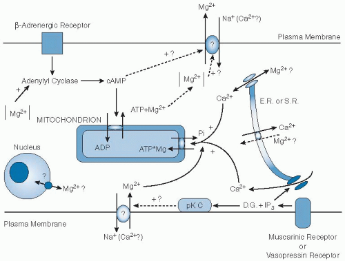
The processes that maintain or modify the relationships between total and ionized internal and external Mg are incompletely understood. Changes in cytosolic Mg2+ regulate some channels (TRPM6 and TRPM7) (24). Mg transport in mammalian cells may be influenced by hormonal and pharmacologic factors (15). Mg2+ efflux was stimulated after short-term exposure of isolated perfused rat heart and liver or thymocytes to α and β-agonists and permeant cAMP (30, 31). Activation of protein kinase C by diacylglycerol or by phorbol esters stimulates Mg2+ influx and does not alter efflux (32). Epidermal growth factor (EGF) has been shown to increase Mg2+ transport into a vascular smooth muscle cell line (33). Insulin and dextrose were found to increase 28Mg uptake by several tissues, including skeletal and cardiac muscle (5, 6). The mechanism of insulin-induced Mg transport is likely the result of an effect on protein kinase C (5, 6). An insulininduced transport of Mg into cells could be one factor responsible for the fall in the serum Mg concentration observed during insulin therapy of diabetic ketoacidosis (34). Investigators have hypothesized that this hormonally regulated Mg uptake system controls intracellular Mg2+ concentration in cellular subcytoplasmic compartments. The Mg2+ concentration in these compartments would then serve to regulate the activity of Mg-sensitive enzymes. An overall schema of cellular Mg homeostasis is shown in Figure 9.1.


Body Homeostasis

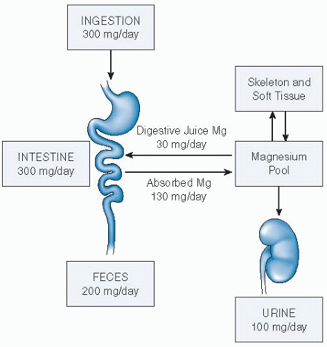
Mineral homeostasis of the individual depends on the amounts ingested, the efficiency of intestinal and renal absorption and excretion, and all other factors affecting them. A schema for human Mg balance is given in Figure 9.2.


Dietary Intake

Mg is widely distributed in plant and animal food sources, but in differing concentrations. Vegetables, fruits, grains, and animal products account for approximately 16% each; dairy products contribute 20% in adolescents and 10% beyond the third decade (35). The 1994 US Department of Agriculture Continuing Survey of Food Intakes by Individuals (CSFII) indicated that the mean daily Mg intake was 323 mg in boys and men and 228 mg in girls and women, findings similar to those of the third National Health and Nutrition Examination Survey (NHANES III). These values fall to less than the current RDA recommendation of approximately 420 mg for boys and men and 320 mg for girls and women (4). Indeed, investigators have suggested that 75% of U.S. residents have a dietary Mg intake lower than the RDA (see the later discussion of Mg requirements and http://ods.od.nih.gov/factsheets/magnesium.asp).


Intestinal Absorption

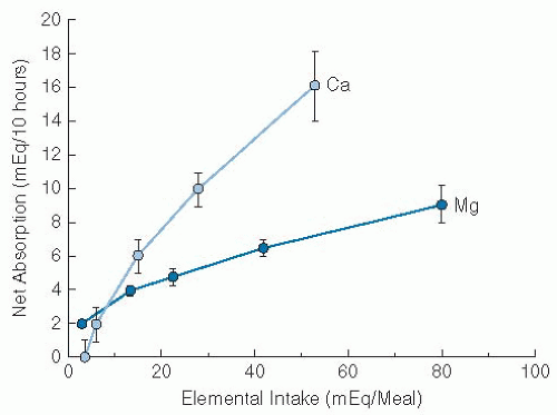
Molecular mechanisms for Mg homeostasis have been reviewed (36). In humans, the primary sites of intestinal Mg absorption are the jejunum and ileum, although absorption can occur at other sites, including the colon (37). Under normal dietary Mg intake, 30% to 40% is absorbed. Following oral ingestion, 28Mg appears in the blood within 1 hour, stabilizes at the rate of 4% to 6%/hour from the second hour to the eighth hour, then decreases rapidly, and ceases at the tenth hour (38). Mg absorption has both a passive paracellular mechanism and an active transport process (Fig. 9.3). The paracellular mechanism depends on a transcellular potential difference generated by Na transport and accounts for approximately 90% of intestinal Mg absorption (37). A Mg-specific transport protein channel, TRPM6 (28), accounts for the remainder
of Mg absorption and may be influenced by certain hormones (39). Absorption of Mg as a function of intake is curvilinear (Fig. 9.3), and this pattern reflects this active saturable process and passive diffusion. Net Mg absorption increases with increasing Mg intake; however, fractional Mg absorption falls. When small amounts of Mg were fed in the form of a standard meal supplemented by varying amounts of Mg (40), fractional absorption fell progressively from approximately 65% to 70% with intake of 7 to 36 mg (0.3 to 1.5 mmol) down to 11% to 14% with intake of 960 to 1000 mg (40 mmol).


Fig. 9.1. Schema of regulation of cellular magnesium (Mg2+) homeostasis in the mammalian cell. The pathways are indicated for cellular Mg2+ release (upper section) and for its uptake (lower section). Stimulated by β-adrenergic agonists, cyclic adenosine monophosphate (cAMP) is increased in the cytosol, which modulates mitochondrial adenine nucleotide translocase and increases the efflux of Mg2+ from the mitochondrion by means of an exchange of one Mg adenosine triphosphate (MgATP) for adenosine diphosphate (ADP). Activation of muscarinic receptors (in cardiac cells) or vasopressin receptors (in the liver) may stimulate an Mg2+ influx mechanism either by decreasing cAMP or by enhancing protein kinase C (pK C) activity by diacylglycerol (D.G.). Vasopressin receptor activation is coupled with production of inositol triphosphate (IP3) from phosphatidylinositol bisphosphate, which induces release of calcium (Ca2+) from the endoplasmic reticulum (E.R.) or the sarcoplasmic reticulum (S.R.). Ca2+ release may be associated with either Mg2+ influx or Mg redistribution in the nucleus or endoplasmic reticulum. Na+, sodium. (Adapted with permission from Romani A, Marfella C, Scarpa A. Cell magnesium transport and homeostasis: role of intracellular compartments. Miner Electrolyte Metab 1993;19:282-9.)

Data on absorption fractions from balance studies using differing diets have been quite variable, ranging from 35% to 70% (41). When free-living adults eating self-selected diets were evaluated periodically over the course of a year, the mean absorptive fraction averaged 21% with an average intake of 323 mg (13.4 mmol) by men and 27% with an average intake of 234 mg (9.75 mmol) by women (42).


Bioavailability

The fractional absorption of ingested Mg by healthy persons is influenced not only by its dietary concentration, but also the presence of dietary components inhibiting or promoting absorption. Long-term balance studies in healthy individuals generally indicate that increasing oral Ca intake does not significantly affect Mg absorption or retention (43). Increased amounts of Mg in the diet have been associated with either decreased Ca absorption (44) or no effect (45). Although a higher Mg intake may not affect intestinal Ca absorption, renal tubular mechanisms may increase Ca excretion (40).

Some reports showed decreased Mg absorption at high intakes of dietary phosphate, whereas others found no consistent effect (46). Increased amounts of absorbable oral Mg have been noted to decrease phosphate absorption, perhaps secondary to formation of insoluble Mg phosphate (40). Reduced absorption of Mg associated with high phosphate intake did not change Mg balance, however, because of associated decreased urinary excretion of Mg (40).

A major increase in zinc intake (from 12 to 142 mg/day) lowered Mg absorption and balance significantly (47). Vitamin B6 depletion induced in young women was associated with a negative Mg balance because of increased urinary excretion (48). The presence of excessive amounts of free fatty acids and oxalate may also impair Mg absorption (49).


Fig. 9.2. Magnesium (Mg) homeostasis in humans. A schematic representation of its metabolic economy indicating (a) its absorption from the alimentary tract, (b) its distribution into bone, and (c) its dependence on the kidney for excretion. Homeostasis depends on the integrity of intestinal and renal absorptive processes. (Adapted with permission from Rude RK. Magnesium homeostasis. In: Bilezikian JB, Raisz L, Rodan G, eds. Principles of Bone Biology. 3rd ed. San Diego: Academic Press, 2008:487-513.)

Increased intakes of dietary fiber have been reported to decrease Mg utilization in humans, presumably by reducing absorption. The introduction of uncontrolled variables, including multiple differences among dietary components in addition to fiber contents, complicates interpretation of the data, however (46). When isolated fiber was added to a basal diet, the effects of fiber itself were negative for dephytinized barley fiber (50) and positive for cellulose (51).


Absorbability of Magnesium Salts

Multiple salts of Mg are available as dietary supplements including oxide, hydroxide, citrate, chloride, gluconate, lactate, and aspartate. The fractional absorption of a salt depends on its solubility in intestinal fluids and the amounts ingested; an amount of 5 mmol (120 mg) of the acetate in gelatin capsules has been found to be an optimal dose in terms of net absorption (40). Absorption of enteric-coated Mg chloride is 67% less than that of the acetate in gelatin capsules (41). In one study, Mg citrate was found to have high solubility, even in water, whereas Mg oxide was poorly soluble, even in acid solution; better absorption of the citrate salt was demonstrated in humans (52). Little difference in absorption has been demonstrated among other salts, however (53). Mg oxide and various salts in large doses act as an osmotic laxative, with resultant diarrhea; the physician faced with a patient who has diarrhea of uncertain origin should consider measuring fecal Mg (45).


Fig. 9.3. Net magnesium (Mg) and calcium (Ca) absorption in healthy humans. The data were obtained under conditions described in reference 39 and in the text. Mean values S.E. are indicated by vertical bars. The absorption data for Mg represent a curved function compatible with a saturable process (at ˜10 mEq/meal in this study) and a linear function reflecting passive diffusion at higher intakes. (Adapted with permission from Fine KD, Santa Ana CA, Porter JL et al. Intestinal absorption of magnesium from foods and supplements. J Clin Invest 1991;88:396-402.)


Regulation of Intestinal Magnesium Absorption

No hormone or factor has been described that regulates intestinal Mg absorption, although several hormones may influence the TRPM6 channel, as discussed earlier. Vitamin D and its active metabolites were shown to increase intestinal Mg absorption in several studies (37). 1,25(OH)2-Vitamin D increases intestinal absorption in normal human subjects and in patients with chronic renal failure (54). In balance studies, vitamin D increased intestinal Mg absorption, but much less than Ca, and mean Mg balance was not affected (54). In patients with impaired Ca absorption resulting from intestinal disease who were given vitamin D, only small increases in Mg absorption were observed compared with Ca (54). Mg was absorbed by individuals with no detectable plasma 1,25(OH)2-vitamin D, and, in contrast to Ca absorption, no significant correlation existed between plasma 1,25(OH)2-vitamin D and Mg absorption (54).


Renal Regulation

Renal Filtration and Tubular Absorption. The kidney is the critical organ regulating Mg homeostasis. Mg handling is a process of filtration and reabsorption. The kidney plays a critical role in excreting the Mg that is not retained for tissue growth or turnover replacement (55). Approximately 10% (roughly 100 mmol or 2400 mg) of total body Mg is normally filtered daily through the glomeruli in the healthy adult; of this, only approximately 5% is excreted in the urine. Approximately 75% of the serum Mg is ultrafiltrable at the glomeruli. The fractional absorption of the filtered load in the various segments of
the nephron is summarized in Figure 9.4. Paracellin-1 (claudin-16) and claudin-19 appear to mediate this transport (27, 28). The distal convoluted tubule reabsorbs 5% to 10% of filtered Mg via an active transcellular pathway. Several proteins may be involved, including the sodium chloride cotransporter (28). TRPM6 is also expressed in the distal tubule. Mutations of TRPM6 result in lower intestinal Mg absorption and renal Mg wasting (27, 28, 29).

Only gold members can continue reading. Log In or Register to continue

Stay updated, free articles. Join our Telegram channel

Jul 27, 2016 | Posted by in PUBLIC HEALTH AND EPIDEMIOLOGY | Comments Off on Magnesium1

Full access? Get Clinical Tree

Get Clinical Tree app for offline access