Patient Story
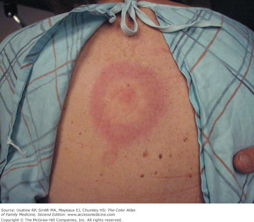
On a warm, summer afternoon a 32-year-old woman presents having had low-grade fevers for 5 days and a rash. On physical examination, the physician notes a large, erythematous, annular patch with central clearing on her back (Figure 218-1). The patient states that the rash has gotten progressively larger during the last 3 days and she has had a recent onset of intermittent joint pain. She does not recall being bitten by an insect. She denies taking medications within the last month and has no known allergies. When asked about recent travel, she admits to a camping trip in eastern Massachusetts, which she returned from 4 days ago. The patient was diagnosed with Lyme borreliosis and started on doxycycline 100 mg twice daily for 14 days. She responded quickly to the antibiotics and never developed the persistent stage of Lyme disease.
Introduction
Lyme disease is an infection caused by the spirochete Borrelia burgdorferi, transmitted via tick bite. Most cases of Lyme disease occur in the northeast United States between April and November. Patients experience flu-like symptoms and may develop the pathognomonic rash, erythema migrans. Lyme disease is prevented by avoiding exposure to the tick vector using insect repellent and protective clothing.
Epidemiology
- In 1977, clusters of patients in Old Lyme, Connecticut, began reporting symptoms originally thought to be juvenile rheumatoid arthritis.1
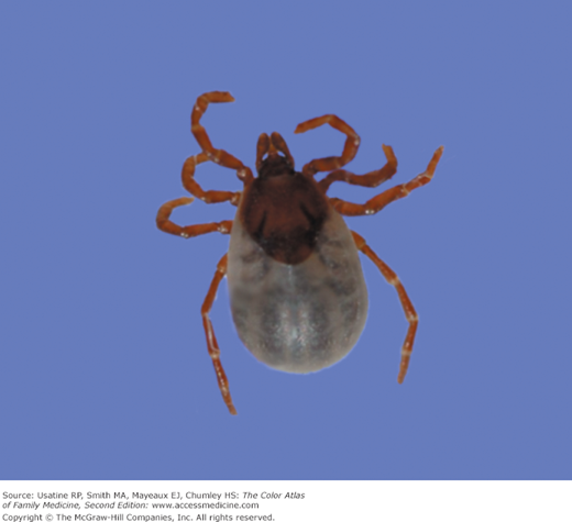
- In 1981, American entomologist, Dr. Willy Burgdorfer, isolated the infectious pathogen responsible for Lyme disease from the midgut of Ixodes scapularis (a.k.a., black-legged deer ticks) (Figure 218-2), which serve as the primary transmission vector in the United States.1
- It was identified as a bacterial spirochete and named B. burgdorferi in honor of its founder.
- Based on Centers for Disease Control and Prevention (CDC) data reported in 2007, Lyme disease (or Lyme borreliosis) is the most common tickborne illness in the United States, with an overall incidence of 7.9 per 100,000 persons.2
- In 2010, 94% of Lyme disease cases were reported from 12 states: Connecticut, Delaware, Maine, Maryland, Massachusetts, Minnesota, New Jersey, New Hampshire, New York, Pennsylvania, Virginia, and Wisconsin.3
- Patients living between Maryland and Maine accounted for 93% of all reported cases in the United States in 2005, with an overall incidence of 31.6 cases for every 100,000 persons.2
- More than 90% of cases report onset between April and November.2
Etiology and Pathophysiology
- B. burgdorferi begins to multiply in the midgut of I. scapularis ticks upon attaching to humans.
- Migration from midgut to salivary glands of ticks requires 24 to 48 hours.
- Prior to this migration, host infection rarely occurs.
- Common hosts include field mice, white-tailed deer, and household pets.
- Ticks must feed on infested hosts in order to infect humans.
- Thirty percent of infected patients do not recall being bitten.4
- Once a human is infected, disease progression is categorized into three stages: localized, disseminated, and persistent.
Diagnosis
Localized (days to weeks)
Erythema migrans (formerly known as erythema chronicum migrans)
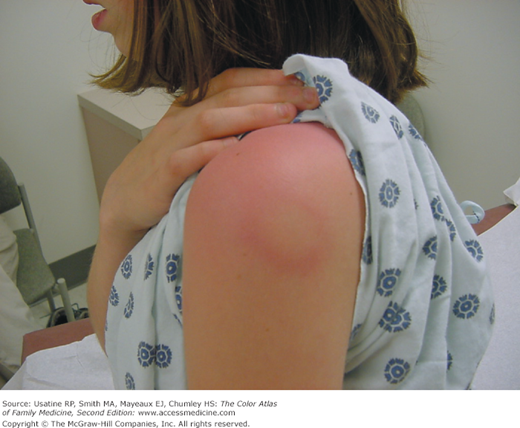
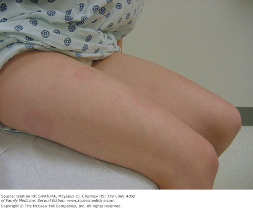
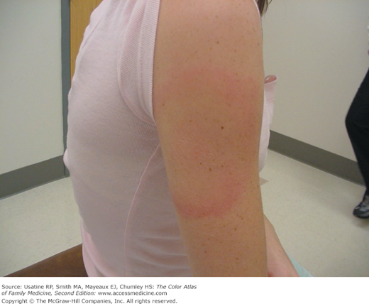
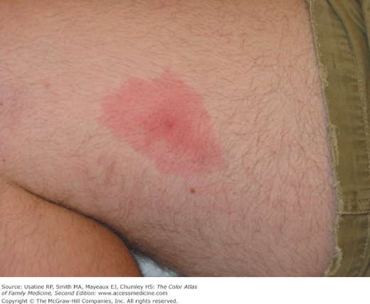
This pathognomonic finding occurs in roughly 68% of Lyme disease cases.4 Described as a “bull’s-eye” eruption (Figures 218-1 and 218-3, 218-4, 281-5, and 218-6), this nonpruritic, maculopapular lesion typically occurs near the site of infection. The erythematous perimeter migrates outward over several days while the central area clears. Multiple lesions in different sites can develop in some individuals (Figures 218-3 and 218-4). Erythema migrans can persist for 2 to 3 weeks if left untreated.
Figure 218-4
Same 11-year-old girl in Figure 218-3 with multiple erythema migrans eruptions on her legs. (Courtesy of Jeremy Golding, MD.)
Flu-like symptoms
Roughly 67% of patients will develop flu-like symptoms that can include fever, myalgias, and lymphadenopathy. Symptoms usually subside within 7 to 10 days.
Stay updated, free articles. Join our Telegram channel

Full access? Get Clinical Tree


