Patient Story
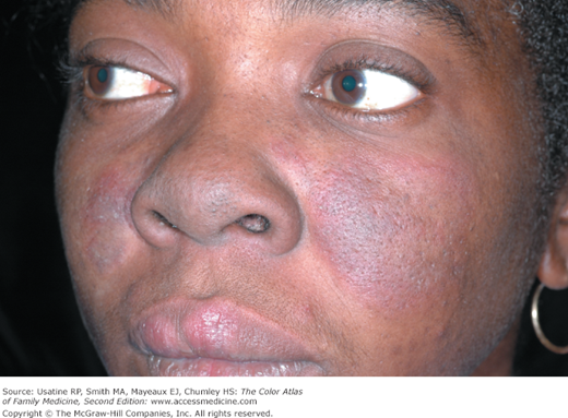
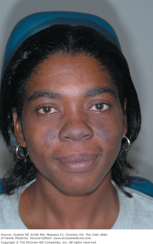
A 39-year-old black woman presented to the clinic with 2 months of swelling of her upper lip and cheeks with new dark spots on her face (Figure 180-1). An antinuclear antibody (ANA) was positive at a 1:80 dilution. A homogeneous nuclear pattern was present as commonly seen in systemic lupus erythematosus (SLE) and drug-induced lupus. The punch biopsy of a facial lesion was consistent with chronic cutaneous lupus erythematosus (discoid lupus). The remainder of her laboratory tests were normal. The patient’s facial lesions did not respond to topical steroids and hence she was started on a short course of systemic steroids. The improvement was seen 3 weeks later (Figure 180-2). Hyperpigmentation remained but erythema, swelling, and pruritus were gone. The patient did not meet criteria for SLE and it is possible to have discoid lupus with a positive ANA. Treatment with hydroxychloroquine was discussed.
Introduction
SLE is a chronic inflammatory disease that can affect many organs of the body including the skin, joints, kidneys, lungs, nervous system, and mucous membranes. Cutaneous lupus can occur in one of three forms: chronic cutaneous (discoid) lupus erythematosus, subacute cutaneous lupus erythematosus, and acute cutaneous lupus erythematosus.
Synonyms
Epidemiology
- In the United States, the prevalence of SLE plus incomplete SLE (disease only partially meeting diagnostic requirements for SLE) is 40 to 50 cases per 100,000 persons.1 It is more common in women and patients with African ancestry.1 Worldwide, the highest SLE prevalences have been reported in Italy, Spain, Martinique, and the United Kingdom Afro-Caribbean population, but it is rarely reported among blacks who live in Africa.2
- Discoid lupus erythematosus (DLE) develops in up to 25% of patients with SLE, but may also occur in the absence of any other clinical feature of SLE.3 Patients with only DLE have a 5% to 10% risk of eventually developing SLE, which tends to follow a mild course.4 DLE lesions usually slowly expand with active inflammation at the periphery, and then to heal, leaving depressed central scars, atrophy, telangiectasias, and hypopigmentation.5 The female-to-male ratio of DLE is 2:1.
Etiology and Pathophysiology
- One proposed mechanism for the etiology of SLE involves the development of autoantibodies that result from a defect in apoptosis. It has been determined that the specific defect involves the “find-me” (and adenosine triphosphate [ATP]/uridine triphosphate [UTP]) or “eat-me” (phosphatidylserine) signals that should be activated when red cell nuclei are extruded. With no apoptosis, the nuclei break down, causing inflammation and the development of autoimmunity.6 Many of the signs and symptoms of lupus erythematosus (LE) are caused by the circulating immune complexes or by the direct effects of antibodies to cells.
- A genetic predisposition for SLE exists. The concordance rate in monozygotic twins is between 25% and 70%. If a mother has SLE, her daughter’s risk of developing the disease is 1:40 and her son’s risk is 1:250.
- The course of SLE is one of intermittent remissions punctuated by disease flares. Organ damage often progresses over time.
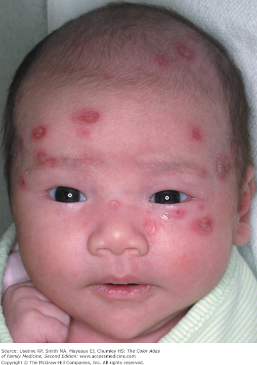
- Rarely, neonates may develop a lupus rush from acquired antibodies through transplacental transmission from mother if she has active SLE (Figure 180-3).
Risk Factors
Diagnosis
- SLE is a chronic, recurrent, potentially fatal inflammatory disorder that can be difficult to diagnose. It is an autoimmune disease involving multiple organ systems that is defined clinically with associated autoantibodies directed against cell nuclei. The disease has no single diagnostic sign or marker. Accurate diagnosis is important because treatment can reduce morbidity and mortality.7
- SLE most often presents with a mixture of constitutional symptoms including fatigue, fever, myalgia, anorexia, nausea, and weight loss. The mean length of time between onset of symptoms and diagnosis is 5 years.
- The disease is characterized by exacerbations and remissions as well as symptoms.
- The diagnosis of SLE is made if four or more of the manifestations mentioned below (and categorized in Table 180-1) are either present, serially or simultaneously, in the patient at the time of presentation or were present in the past. If two to three manifestations are present, some clinicians refer to the syndrome as “incomplete lupus.”8
- Arthralgias, which are often the initial complaint, are usually disproportionate to physical findings. The polyarthritis is symmetric, nonerosive, and usually nondeforming. In longstanding disease, rheumatoid-like deformities with swan-neck fingers are commonly seen.
- A malar or butterfly rash is fixed erythema over the cheeks and bridge of the nose sparing the nasolabial folds (Figures 180-2, 180-4, and 180-5). It may also involve the chin and ears. More severe malar rashes may cause severe atrophy, scarring, and hypopigmentation (Figure 180-5).
- Rash associated with photosensitivity to UV light.
- A discoid rash consisting of erythematosus raised patches with adherent keratotic scaling and follicular plugging. Atrophic scarring may occur in older lesions.
- Ulcers (usually painless) in the nose, mouth, or vagina are frequent complaints.
- Pleuritis as evidenced by a convincing history of pleuritic pain or rub or evidence of pleural effusion.
- Pericarditis as documented by ECG, rub, or evidence of pericardial effusion.
- Renal disorder such as cellular casts or persistent proteinuria greater than 0.5 g/day or greater than 3+ if quantitation not performed.
- Central nervous system (CNS) symptoms ranging from mild cognitive dysfunction to psychosis or seizures. Any region of CNS can be involved. Intractable headaches and difficulties with memory and reasoning are the most common features of neurologic disease in lupus patients.
- Hematologic disorders such as hemolytic anemia, leukopenia (<4000/mm3 total on two or more occasions), lymphopenia (<1500/mm3 on two or more occasions), or thrombocytopenia (<100,000/mm3 in the absence of precipitating drugs).
- GI symptoms may include abdominal pain, diarrhea, and vomiting. Intestinal perforation and vasculitis are important diagnoses to exclude.
- Vasculitis (Figures 180-6,180-7, and 180-8) can be severe and can include retinal vasculitis.
- Immunologic disorders such as a positive antiphospholipid antibody, anti-DNA, anti-Smith antigen, or a false-positive serologic test for syphilis (known to be positive for at least 6 months and confirmed by a negative treponema specific test).
- An abnormal titer of ANA at any point in time and in the absence of drugs associated with “drug-induced lupus.”
- Arthralgias, which are often the initial complaint, are usually disproportionate to physical findings. The polyarthritis is symmetric, nonerosive, and usually nondeforming. In longstanding disease, rheumatoid-like deformities with swan-neck fingers are commonly seen.
Criterion | Definition |
|---|---|
1. Malar rash | Fixed erythema, flat or raised, over the malar eminences, tending to spare the nasolabial folds |
2. Discoid rash | Erythematosus-raised patches with adherent keratotic scaling and follicular plugging and later atrophic scarring |
3. Photosensitivity | Skin rash as a result of unusual reaction to sunlight, by history or physician observation |
4. Oral ulcers | Oral or nasopharyngeal ulceration, usually painless, observed by a physician |
5. Arthritis | Nonerosive arthritis involving 2 or more peripheral joints, characterized by tenderness, swelling, or effusion |
6. Serositis | Pleuritis—convincing history of pleuritic pain or rub heard by a physician or evidence of pleural effusion or pericarditis documented by ECG, rub, or evidence of pericardial effusion |
7. Renal disorder | Persistent proteinuria greater than 0.5 g/day or greater than 3+ if quantitation not performed or red cell, hemoglobin, granular, tubular, or mixed cellular casts |
8. Neurologic disorder | Seizures or psychosis—in the absence of offending drugs or known metabolic derangements (uremia, ketoacidosis, or electrolyte imbalance) |
9. Hematologic disorder | Hemolytic anemia with reticulocytosis or leukopenia (<4,000/mm3 on 2 or more occasions) or lymphopenia (<1,500/mm3 on 2 or more occasions) or thrombocytopenia (<100,000/mm3) in the absence of offending drugs |
10. Immunologic disorders | Positive antiphospholipid antibody or anti-DNA antibody to native DNA in abnormal titer or anti-Smith antibody—presence of antibody to Smith nuclear antigen or false-positive serologic test for syphilis known to be positive for at least 6 months and confirmed by Treponema pallidum immobilization or fluorescent treponemal antibody absorption test |
11. Antinuclear antibody | An abnormal titer of antinuclear antibody by immunofluorescence or an equivalent assay at any point in time and in the absence of drugs known to be associated with “drug-induced lupus” syndrome |


