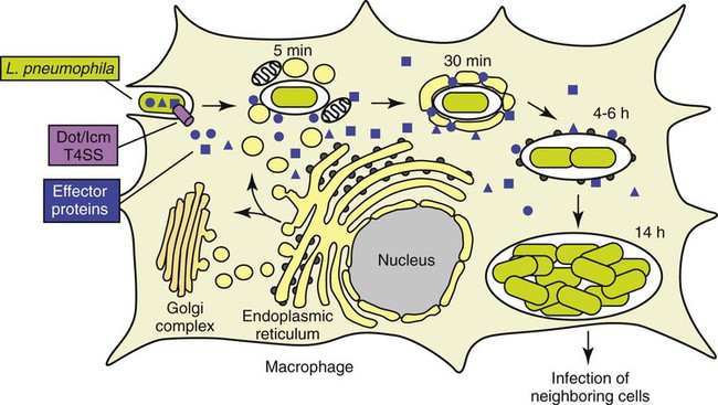
Legionella


Legionella


This chapter addresses organisms that will not grow on routine primary plating media and belong to the genus Legionella. Legionella belongs to the family Legionellaceae and includes a single genus, Legionella, comprising approximately 52 species. Legionella pneumophila is the causative agent of Legionnaires’ disease, a febrile and pneumonic illness with numerous clinical presentations. Legionella was discovered in 1976 by scientists at the Centers for Disease Control and Prevention (CDC) who were investigating an epidemic of pneumonia among Pennsylvania State American Legion members attending a convention in Philadelphia. There is retrospective serologic evidence of Legionella infection as far back as 1947. Bacteria resembling Legionella that are capable of living in amoebae have been designated as Legionella-like amoebal pathogens (LLAPs).


General Characteristics


All Legionella spp. are mesophilic (20° to 45° C), obligately aerobic, faintly staining, thin, gram-negative fastidious bacilli that require a medium supplemented with iron and L-cysteine, and buffered to pH 6.9 for optimum growth. The organisms utilize protein for energy generation rather than carbohydrates. The overwhelming majority of Legionella spp. are motile. As of this writing, more than 52 species belong to this genus. Nevertheless, the organism Legionella pneumophila predominates as a human pathogen within the genus and consists of 16 serotypes. In approximately decreasing order of clinical importance are L. pneumophila serotype 1 (about 70% to 90% of the cases of Legionnaires’ disease), L. pneumophila serotype 6, L. micdadei, L. dumoffii, L. anisa, and L. feeleii. Of note, many species of Legionella have only been isolated from the environment or recorded as individual cases. To date, 20 species of Legionella are documented as human pathogens in addition to L. pneumophila. Box 35-1 is an abbreviated list of some of the species of Legionella.


Epidemiology


Legionellae are ubiquitous and widely distributed in the environment. As a result, most individuals are exposed to Legionella spp.; however, few develop symptoms. In nature, legionellae are found primarily in aquatic habitats and thrive at warmer temperatures; these bacteria are capable of surviving extreme ranges of environmental conditions for long periods; studies have shown that L. pneumophila can survive for up to 14 months in water with only a slight loss in viability. Legionella spp. have been isolated from the majority of natural water sources investigated, including lakes, rivers, and marine waters, as well as moist soil. Organisms are also widely distributed in man-made facilities, including air-conditioning ducts and cooling towers; potable water; large, warm-water plumbing systems; humidifiers; whirlpools; and technical-medical equipment in hospitals.


Legionella infections are acquired exclusively from environmental sources; no person-to-person spread has been documented. Inhalation of infectious aerosols (1 to 5 μm in diameter) is considered the primary means of transmission. Exposure to these aerosols can occur in the workplace or in industrial or nosocomial settings; for example, nebulizer’s filled with tap water and showers have been implicated. Infection is acquired through the inhalation of aerosols or microaspiration. Legionnaires’ disease occurs in sporadic, endemic, and epidemic forms. The incidence of disease varies greatly and appears to depend on the geographic area, but it is estimated that Legionella spp. cause less than 1% to 5% of cases of pneumonia.


Pathogenesis and Spectrum of Disease


Legionella spp. can infect and multiply within some species of free-living amoebae (Hartmannella, Acanthamoeba, and Naegleria spp.), as well as within Tetrahymena spp., a ciliated protozoa, or within biofilms (well-organized microcolonies of bacteria usually enclosed in polymer matrices that are separated by water channels that remove wastes and deliver nutrients). This contributes to the organism’s survival in the environment. In addition, L. pneumophila exists in two well-defined, morphologically distinct forms in Hela cells: (1) a highly differentiated, cystlike form that is highly infectious, metabolically dormant, and resistant to antibiotics and detergent-mediated lysis and (2) a replicative intracellular form that is ultrastructurally similar to agar-grown bacteria. The existence of this cystlike form may account for the ability of L. pneumophila to survive for long periods between hosts (amoebae or humans).


Although the exact mechanisms by which L. pneumophila causes disease are not totally delineated, its ability to avoid destruction by the host’s phagocytic cells plays a significant role in the disease process. L. pneumophila is considered a facultative intracellular pathogen. Following infection, organisms are taken up by phagocytosis primarily in alveolar macrophages, where they survive and replicate within a specialized, membrane-bound vacuole by resisting acidification and evading fusion with lysosomes; it is still unknown how Legionella prevent vacuole acidification. Following replication, the organisms will kill the phagocyte releasing them into the lungs and will again be phagocytized by a mononuclear cell, and multiplication of the organism will increase.


The sequestering of legionellae within macrophages also makes it difficult to deliver and accumulate effective antimicrobials. Of significance, studies have shown that although certain antimicrobials can penetrate the macrophage and inhibit bacterial multiplication, L. pneumophila is not killed and, when drugs are removed, the organism resumes replicating. Therefore, a competent cell-mediated immune response is also important for recovery from Legionella infections. Humoral immunity appears to play an insignificant role in the defense against this organism.


In eukaryotic cells, most proteins secreted or transported inside vesicles to other cellular compartments are synthesized at the endoplasmic reticulum (ER) (Figure 35-1). Many bacterial pathogens use secretion systems as a part of how they cause disease. L. pneumophila possesses genes that are able to “trick” eukaryotic cells into transporting them to the endoplasmic reticulum; these virulence genes are called dot (defective organelle trafficking) or icm (intracellular multiplication). This dot/icm secretion system in L. pneumophila consists of 23 genes and is a type IV secretion system. Bacterial type IV secretion systems are bacterial devices that deliver macromolecules such as proteins across and into cells. After entry but before bacterial replication, L. pneumophila, residing in a membrane-bound vacuole, is surrounded by a ribosome-studded membrane derived from the host cell’s ER and mitochondria. Thus, by exploiting host cell functions, L. pneumophila is able to gain access to the lumen of the ER, which supports its survival and replication where the environment is rich in peptides. A second type II secretion system has also been implicated in the virulence of some strains of Legionella. The type II secretion system carries numerous genes for enzymatic degradation including lipases, proteinases, and a number of novel proteins. Mutations within the type II secretion system results in decreased infectivity of the organism. A number of additional bacterial factors have also been identified as crucial for intracellular infection; some of these are listed in Box 35-2.


image
FIGURE 35-1
(Modified from 2009annualreport.nichd.nih.gov/ump.html.)

Finally, several cellular components and extracellular products of L. pneumophila, such as an extracellular cytotoxin that impairs the ability of phagocytic cells to use oxygen and various enzymes (e.g., phospholipase C), have been purified and proposed as virulence factors. However, their exact role in the pathogenesis of Legionella infections is not completely clear.


Legionella spp. are associated with a spectrum of clinical presentations, ranging from asymptomatic infection to severe, life-threatening diseases. Serologic evidence exists for the presence of asymptomatic disease, because many healthy people surveyed possess antibodies to Legionella spp. Table 35-1 provides a more detailed description of the following three primary clinical manifestations:


Individuals at risk for pneumonia are those who are immunocompromised, older than age 60, or heavy smokers. The clinical manifestations following infection with a particular species are primarily caused by differences in the host’s immune response and perhaps by inoculum size; the same Legionella sp. gives rise to different expressions of disease in different individuals.


There are a number of bacteria that grow only within amoebae and are closely related phylogenetically based on 16S rRNA gene sequencing to Legionella species; these organisms are referred to as “Legionella-like amoeba pathogens” (LLAPs). Several LLAPs have been assigned to the Legionella genus. One LLAP has been isolated from the sputum of a patient with pneumonia after the specimen was incubated with the amoeba Acanthamoeba polyphaga. Serologic surveys of patients with community-acquired pneumonia suggest LLAPs may be occasional human pathogens.


Laboratory Diagnosis


Specimen Collection and Transport


Specimens from which Legionella can be isolated include respiratory tract secretions of all types, including expectorated sputum, additional lower respiratory specimens, and pleural fluid; other sterile body fluids, such as blood; and lung, transbronchial, or other biopsy material. Because sputum from patients with Legionnaires’ disease is usually nonpurulent and may appear bloody or watery, the grading system used for screening sputum for routine cultures is not applicable. Patients with Legionnaires’ disease usually have detectable numbers of organisms in their respiratory secretions, even for some time after antibiotic therapy has been initiated. If the disease is present, the initial specimen is often likely to be positive. However, additional specimens should be processed if the first specimen is negative and suspicion of the disease persists. Pleural fluid has not yielded many positive cultures in studies performed in several laboratories, but it may contain organisms. Urine for antigen collection should be collected in a sterile container. The sample should be transported to the laboratory and refrigerated if a delay in processing occurs. Specimens should be transported without holding media, buffers, or saline, which may inhibit the growth of Legionella. The organisms are hardy and are best preserved by maintaining specimens in a small, tightly closed container to prevent desiccation and transporting them to the laboratory within 30 minutes of collection. If a longer delay is anticipated, specimens should be refrigerated. If one cannot ensure that specimens will remain moist, 1 mL of sterile broth may be added.


Specimen Processing


All specimens for Legionella culture should be handled and processed in a class II biologic safety cabinet (BSC). When specimens from nonsterile body sites are submitted for culture, selective media or treatment of the specimen to reduce the numbers of contaminating organisms is proposed. Brief treatment of sputum specimens with hydrochloric acid before culture has been shown to enhance the recovery of legionellae. However, this technique is time consuming and is only recommended on specimens from patients with cystic fibrosis. Respiratory secretions may be held for up to 48 hours at 5° C before culture; if culturing is delayed longer, then the specimen may be frozen.


Tissues are homogenized before smears and cultures are performed, and clear, sterile body fluids are centrifuged for 30 minutes at 4000× g. The sediment is then vortexed and used for culture and smear preparation. Blood for culture of Legionella may be processed with the lysis-centrifugation tube system (Isolator; Wampole Laboratories, Cranbury, New Jersey) and plated directly to buffered charcoal-yeast extract (BCYE) agar. Specimens collected by bronchoalveolar lavage are quite dilute and therefore should be concentrated at least tenfold by centrifugation before culturing.

Stay updated, free articles. Join our Telegram channel

Aug 25, 2016 | Posted by in MICROBIOLOGY | Comments Off on Legionella

Full access? Get Clinical Tree

Get Clinical Tree app for offline access