Patient Story
A 71-year-old woman presented with a rapidly growing lesion on her face over the past 4 months (Figure 167-1). The lesion had features of a basal cell carcinoma with a pearly border and telangiectasias (Figure 167-2). Also the central crater with keratin gave it the appearance of a keratoacanthoma (KA). A shave biopsy was performed and the pathology showed squamous cell carcinoma (SCC)-KA type. A full elliptical excision with 4-mm margins was then performed.
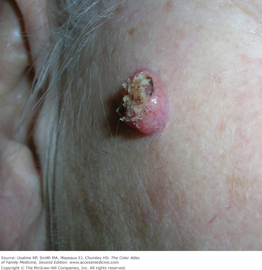
Figure 167-2
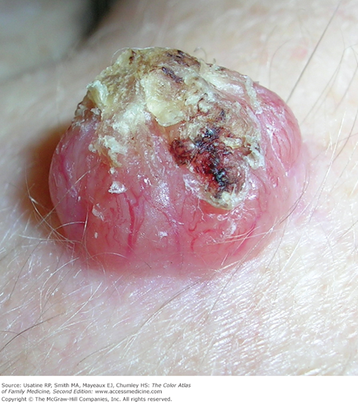
Close-up of the keratoacanthoma with telangiectasias and a central keratin core on the face of the woman in Figure 167-1. (Courtesy of Richard P. Usatine, MD.)
Introduction
The KA is a unique epidermal tumor characterized by rapid, abundant growth and a spontaneous resolution, with the classic presentation in middle-aged, light-skinned individuals in hair-bearing, sun-exposed areas. In the late 1940s, Freudenthal of Wroclaw coined the term keratoacanthoma, owing to the considerable acanthosis observed in the tumor. Controversies have arisen since the 1950s about the real nature of the tumor; some KAs may metastasize, and there is debate over the relationship to SCC.1,2 Many dermatopathologists now classify this tumor as a subtype of SCC.
Synonyms
- Keratocarcinoma.1
- Molluscum sebaceum.
- Molluscum pseudocarcinomatosum.
- Cutaneous sebaceous neoplasm.
- Self-healing squamous epithelioma.
- Intracutaneous cornifying epithelioma.
- Idiopathic cutaneous pseudoepitheliomatous hyperplasia.
- Verrugoma.
Epidemiology
Etiology and Pathophysiology
- KAs share features such as infiltration and cytologic atypia with SCCs.
- KAs have been reported to metastasize.
- KA is considered to be a variant of SCC, called SCC-KA type.
- Histologic criteria are not sensitive enough to discriminate reliably between KA and SCC.4


