• Increased fibroblasts, lymphocytes, and mast cells usually present
Top Differential Diagnoses
• Hypertrophic scar
Lacks characteristic hyalinized collagen bundles of keloid
• Nodular fasciitis
May rarely show focal keloidal collagen but should have background of typical loose stroma
• Desmoplastic melanoma
Rarely may enter differential diagnosis if no history of trauma or previous biopsy/surgery
• Dermatofibroma
• Keloidal atypical fibroxanthoma
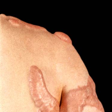
Clinical Photograph of Multiple Keloids Clinical photograph shows multiple keloids on the shoulder and upper arm of this patient.
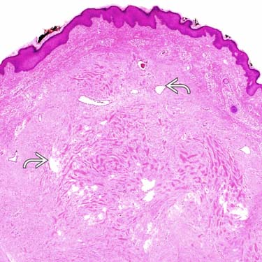
Scanning Magnification of Keloid Scanning magnification of a keloid shows a polypoid skin lesion with dense dermal collagen. Note the mild epidermal hyperplasia and telangiectatic vessels .
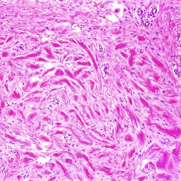
High Magnification of Keloidal Collagen High-power examination of a keloid shows a proliferation of thickened, hyalinized eosinophilic collagen bundles with increased numbers of stromal fibroblasts. Collagen bundles are randomly oriented and unevenly distributed.
Keloid With Telangiectasia Superficial portion of keloid shows telangiectatic vessels surrounded by thickened collagen bundles.
TERMINOLOGY
Synonyms
• Scar with keloidal collagen
Definitions
• Scar with prominent thickened and eosinophilic bundles of collagen extending beyond original wound
ETIOLOGY/PATHOGENESIS
Unknown, Possibly Genetic
• Fibroblasts from keloids show decreased apoptosis
• Many cytokines implicated in stimulating fibroblasts, including TGF-β, heat shock proteins, and IL-15
CLINICAL ISSUES
Epidemiology
• Age
Most common in patients < 30 yr
• Ethnicity
More common in patients of African lineage; least common in Caucasians
Site
• Earlobe is most common site
Typically follows ear piercing or other trauma
Presentation
• Nodule/mass lesion is most common
Only gold members can continue reading. Log In or Register to continue


 .
.
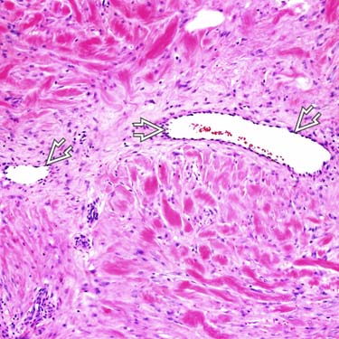
 surrounded by thickened collagen bundles.


