Histologically identical to kaposiform hemangioendothelioma, also occurs in children
Likely in same spectrum of tumors
• Kaposi sarcoma
Older and immunocompromised patients
Positive for HHV8
• Spindle cell hemangioma
Adults, usually distal extremities
Often partially intravascular
Spindled cells, blister cells, and ectatic vascular spaces
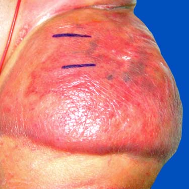
Kaposiform Hemangioendothelioma in Groin of Infant Clinical photograph shows a large inguinal tumor in an infant. More superficial tumors typically present as an erythematous or violaceous mass.
Kaposiform Hemangioendothelioma at Low Magnification Histologic examination at low power of kaposiform hemangioendothelioma demonstrates irregular nodules of spindled cells in the dermis as well as ectatic vessels at periphery of nodules of spindled cells .
Glomeruloid Area in Kaposiform Hemangioendothelioma In some areas, the spindled tumor cells can have a somewhat swirling growth pattern, imparting a glomeruloid appearance.
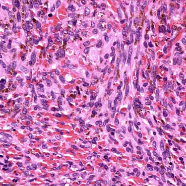
Kaposiform Hemangioendothelioma: Kaposi Sarcoma-Like Area The tumor cells have uniform hyperchromatic spindled nuclei and are arranged in short fascicles with slit-like vascular lumina.
ETIOLOGY/PATHOGENESIS
Developmental Anomaly
• ~ 1/2 of kaposiform hemangioendothelioma cases present in 1st year of life
CLINICAL ISSUES
Epidemiology
• Age
Majority present in infancy to teenage years
Presentation
• Painful or painless mass
Presents as superficial or deep mass
Cutaneous lesions present as violaceous plaques
Only gold members can continue reading. Log In or Register to continue

 Highest risk for coagulopathy in retroperitoneal and intrathoracic tumors, but also seen in association with large cutaneous tumors


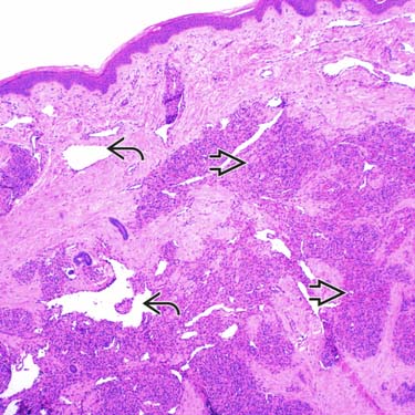
 of spindled cells in the dermis as well as ectatic vessels at periphery of nodules of spindled cells
  .
.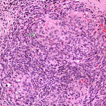
 appearance.