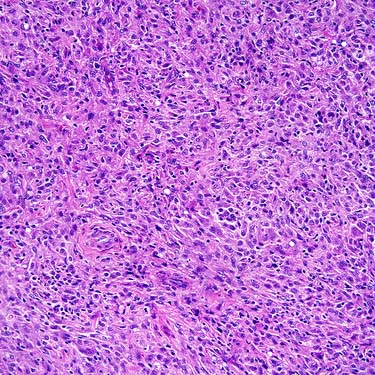
Juvenile Xanthogranuloma This is a typical appearance of a juvenile xanthogranuloma removed from the face of a small boy. The lesion is uniform and cellular and proliferates in the dermis with no grenz zone.
Juvenile Xanthogranuloma at High Magnification A cellular example at high magnification shows numerous mononucleated histiocytes associated with an inflammatory background composed mostly of mononuclear cells.
Juvenile Xanthogranuloma: Touton Cells Many Touton giant cells can be seen in this juvenile xanthogranuloma. The background cells are spindled to ovoid with eosinophilic cytoplasm, which shows only minimal lipid in this case.
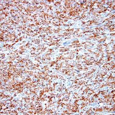
CD68 Staining in Juvenile Xanthogranuloma Essentially all examples of juvenile xanthogranuloma are reactive with CD68 (shown) and factor XIIIa by immunohistochemistry, but are negative for S100 and CD1a.
TERMINOLOGY
Abbreviations
• Juvenile xanthogranuloma (JXG)
Synonyms
• Nevoxanthoendothelioma
Definitions
• Stable or regressing histiocytic lesion that usually occurs in childhood
Form of non-Langerhans histiocytosis
CLINICAL ISSUES
Epidemiology
• Incidence
Rare
• Age
Majority in individuals under 3 years
– Visceral examples almost exclusively in infants and children
13-30% in older children and adults
• Sex
Slight male predominance
Presentation
• Solitary cutaneous lesion in majority of cases
Head and neck > trunk > extremities
• Up to 10% of patients with multiple cutaneous lesions
• Up to 5% of patients with visceral-systemic disease
Treatment
• Simple excision
• Chemotherapy administered to rare patients with systemic disease
Prognosis
• Usually excellent
Most lesions regress or stabilize (including large visceral ones)
Rare deaths associated with multiorgan disease
MICROSCOPIC
Histologic Features
• Mononuclear cells typically predominate
• Multinucleated cells ± Touton features
• Spindle cells (typically minor component)
Variable finely vacuolated cytoplasm
Often lightly eosinophilic
• Variable lipid and foamy histiocytes
Minimal lipid in early lesions
Only gold members can continue reading. Log In or Register to continue
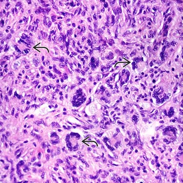
 can be seen in this juvenile xanthogranuloma. The background cells are spindled to ovoid with eosinophilic cytoplasm, which shows only minimal lipid in this case.