Chapter 17
Introduction to genetics
![]() Animations
Animations
17.2 Sex determination 438
17.3 Complementary base pairing of DNA 440
17.4 Transcription 441
17.5 Translation 441
17.6 Crossing over 443
17.7 Hereditary traits 444
17.8 Punnett square 444
17.9 Sex-linked traits 444
The Human Genome project, an international collaboration, was initiated in 1990. Its aim was to identify and sequence every gene on every chromosome, and to be able to draw a map of each chromosome, showing the position of all its genes. It has yielded a great deal of important information regarding human genetic disorders.
At the end of the chapter, the effects of ageing on chromosomes, cell division and heredity are considered, and this is followed by a section on some common genetic abnormalities.
Chromosomes, genes and DNA
Chromosomes
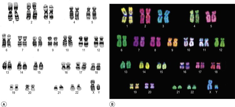
Nearly every body cell contains, within its nucleus, an identical copy of the entire complement of the individual’s genetic material. Two important exceptions are red blood cells (which have no nucleus) and the gametes or sex cells. In a resting cell, the chromatin (genetic material, see Fig. 3.8) is diffuse and hard to see under the microscope, but when the cell prepares to divide, it is collected into highly visible, compact, sausage-shaped structures called chromosomes. Each chromosome is one of a pair, one inherited from the mother and one from the father, so the human cell has 46 chromosomes that can be arranged as 23 pairs. A cell with 23 pairs of chromosomes is termed diploid. Gametes (spermatozoa and ova) with only half of the normal complement, i.e. 23 chromosomes instead of 46, are described as haploid. Chromosomes belonging to the same pair are called homologous chromosomes. The complete set of chromosomes from a cell is its karyotype (Fig. 17.1). ![]() 17.1
17.1

Figure 17.1 Chromosomal complement (karyotype) of a normal human male, showing 22 pairs of autosomes and sex chromosomes (XY, pair 23).
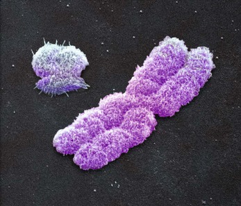
Each pair of chromosomes is numbered, the largest pair being no. 1. The first 22 pairs are collectively known as autosomes, and the chromosomes of each pair contain the same amount of genetic material. The chromosomes of pair 23 are called the sex chromosomes (Fig. 17.2) because they determine the individual’s gender. Unlike autosomes, these two chromosomes are not necessarily the same size; the Y chromosome is much shorter than the X and is carried only by males. A child inheriting two X chromosomes (XX), one from each parent, is female, and a child inheriting an X from his mother and a Y from his father (XY) is male. ![]() 17.2
17.2

Figure 17.2 Coloured scanning electron micrograph of replicated human sex chromosomes: Y upper left, X centre.
Each end of the chromosome is capped with a length of DNA called a telomere, which seals the chromosome and is structurally essential. During replication, the telomere is shortened, which would damage the chromosome, and so it is repaired with an enzyme called telomerase. Reduced telomerase activity with age is related to cell senescence (p. 445).
Genes
Along the length of the chromosomes are the genes. Each gene contains information in code that allows the cell to make (almost always) a specific protein, the so-called gene product. Each gene codes for one specific protein, and research puts the number of genes in the human genome at between 25 000 and 30 000.
Genes normally exist in pairs, because the gene on one chromosome is matched at the equivalent site (locus) on the other chromosome of the pair.
DNA
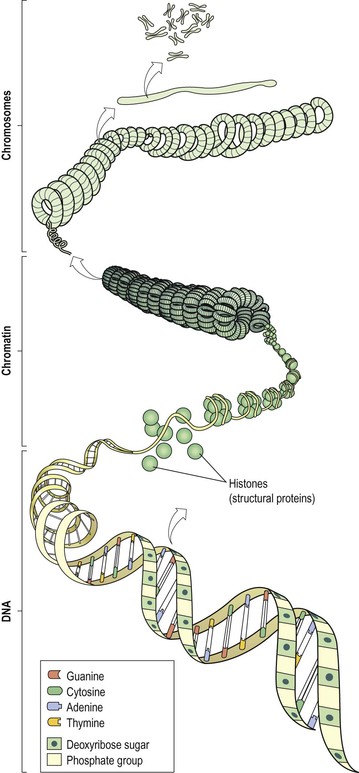
Genes are composed mainly of very long strands of DNA; the total length of DNA in each cell is about a metre. Because this is packaged into chromosomes, which are micrometres (10−6 m) long, this means that the DNA must be tightly wrapped up to condense it into such a small space.
DNA is a double-stranded molecule, made up of two chains of nucleotides. Nucleotides consist of three subunits:
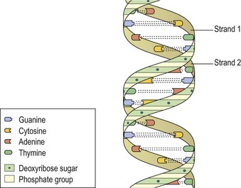
The DNA molecule is sometimes likened to a twisted ladder, with the uprights formed by alternating chains of sugar and phosphate units (Fig. 17.3). In DNA, the sugar is deoxyribose, thus DNA. The bases are linked to the sugars, and each base binds to another base on the other sugar/phosphate chain, forming the rungs of the ladder. The two chains are twisted around one another, giving a double helix (twisted ladder) arrangement. The double helix itself is further twisted and wrapped in a highly organised way around structural proteins called histones, which are important in maintaining the heavily coiled three-dimensional shape of the DNA. The term given to the DNA–histone material is chromatin. The chromatin is supercoiled and packaged into the chromosomes shortly before the cell divides (Fig. 17.4).
The genetic code
DNA carries a huge amount of information that determines all biological activities of an organism, and which is transmitted from one generation to the next. The key to how this information is kept is found in the bases within DNA. There are four bases:
They are arranged in a precise order along the DNA molecule, making a base code that can be read when protein synthesis is required. Each base along one strand of DNA pairs with a base on the other strand in a precise and predictable way. This is known as complementary base pairing. Adenine always pairs with thymine (and vice versa), and cytosine and guanine always go together. The bases on opposite strands run down the middle of the helix and bind to one another with hydrogen bonds (Fig. 17.3). ![]() 17.3
17.3
Mitochondrial DNA
Each body cell has, on average, 5000 mitochondria (p. 33) that hold a quantity of DNA (mitochondrial DNA), which codes, for example, for enzymes important in energy production. This DNA is passed from one generation to another via the ovum (p. 463), so the offspring’s complement of mitochondrial DNA is inherited from the mother. Certain rare inherited disorders that arise from faulty mitochondrial DNA are therefore passed through generations via the maternal line.
Mutation
Mutation means an inheritable alteration in the normal genetic make-up of a cell. Most mutations occur spontaneously, because of the countless millions of DNA replications and cell divisions that occur normally throughout life. Others may be caused by external factors, such as X-rays, ultraviolet rays or exposure to certain chemicals. Any factor capable of mutating DNA is called a mutagen (p. 55). Most mutations are immediately repaired by an army of enzymes present in the cell nucleus, and therefore cause no permanent problems.
Sometimes the mutation is lethal, because it disrupts some essential cellular function, causing cell death and the mutation is destroyed along with the cell. Often, the mutated cell is detected by immune cells and destroyed because it is abnormal (p. 379). Other mutations do not kill the cell but alter its function in some way that may cause disease, e.g. in cancer (p. 55). A persistent mutation in the genome that has not led to cell death can be passed from parent to child and may cause inherited disease, e.g. phenylketonuria (p. 446) or cystic fibrosis (p. 266).


