Introduction to Diseases with Monoclonal Immunoglobulin Deposits
Robert B. Colvin, MD
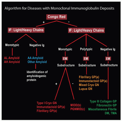 Diagnosis of diseases with monoclonal immunoglobulin deposits &/or substructure by EM is facilitated by following the path in this algorithm, which begins with whether the deposits are Congo red positive, followed by immunofluorescence (IF) for monotypic immunoglobulin chains (single light &/or heavy chain), equal staining of light chains (polyclonal) or no immunoglobulin staining. Electron microscopy is then used for assessment of the substructure. Immunotactoid and fibrillary GP sometimes have monotypic light chain (a). MIDD has either light chain, heavy chain, or both (b) and has no organized substructure. PGNMID usually, but not always, has amorphous deposits (c). |
ETIOLOGY/PATHOGENESIS
Monoclonal Immunoglobulin
May be produced by malignant lymphoid or plasma cell neoplasm
Reactive (Benign Monoclonal Proliferation)
Some represent “monoclonal gammopathy of undetermined significance” (MGUS) without identifiable underlying neoplasm at time of renal biopsy
Some cases never have an identified neoplastic proliferation
These appear to be clonal, but not malignant
APPROACH
Light Microscopy
Quite variable appearances
Mimic membranoproliferative GN, acute GN, membranous GN (MIDD, type I cryo, PGNMID)
Mimic diabetes with nodular mesangial glomerulopathy (MIDD)
Tubulointerstitial disease may predominate (MIDD, cast nephropathy, light chain proximal tubulopathy)
Special Stains
Congo red stain essential to distinguish amyloidosis
Amyloidosis can be due to monoclonal immunoglobulin or other proteins
Thicker sections more sensitive
High-quality polarizing microscope more sensitive
Immunofluorescence
Stains for light chains essential to distinguish this group of diseases
Occasional forms show only heavy chain deposits
Some monotypic light chains truncated & stain poorly
Electron Microscopy
Stay updated, free articles. Join our Telegram channel

Full access? Get Clinical Tree


