and Natalia Buza1
(1)
Department of Pathology, Yale University School of Medicine, New Haven, CT, USA
Keywords
Gestational trophoblastic diseaseComplete hydatidiform molePartial hydatidiform moleInvasive hydatidiform moleGestational choriocarcinomaPlacental site trophoblastic tumorEpithelioid trophoblastic tumorExaggerated placental site reactionPlacental site noduleIntroduction
This chapter covers the frozen section evaluation of uterine contents for confirmation of intrauterine pregnancy and gestational trophoblastic disease (GTD). The latter encompasses a spectrum of proliferative disorders of placental trophoblast ranging from nonneoplastic hydatidiform moles (partial and complete mole) to malignant trophoblastic tumors [1] (Table 6.1). The most aggressive form of trophoblastic neoplasia, gestational choriocarcinoma, has become uncommon nowadays. Two distinct tumors of the intermediate trophoblast (placental site trophoblastic tumor and epithelioid trophoblastic tumor) are rare but frequently pose a diagnostic challenge when encountered. In addition, two reactive conditions (exaggerated placental site reaction and placental site nodule) are also classified within GTD due to their potential misdiagnosis as neoplastic processes.
Table 6.1
2014 World Health Organization classification of gestational trophoblastic disease
Classification | Subtypes | Trophoblastic cell of origin |
|---|---|---|
Hydatidiform mole | Complete mole | Villous trophoblast |
Partial mole | Villous trophoblast | |
Invasive mole | Villous trophoblast | |
Trophoblastic tumors | Gestational choriocarcinoma | Villous trophoblast |
Placental site trophoblastic tumor | Implantation site intermediate trophoblast | |
Epithelioid trophoblastic tumor | Chorionic intermediate trophoblast | |
Nonneoplastic lesions | Exaggerated placental site reaction | Implantation site intermediate trophoblast |
Placental site nodule | Chorionic intermediate trophoblast | |
Abnormal villous lesions | Various non-molar lesions histologically simulating partial mole | Villous trophoblast |
Although rarely requested, possible scenarios for frozen section evaluation include assessment for the presence of persistent trophoblastic disease, particularly invasive mole and metastatic trophoblastic lesions (metastatic mole) and diagnosis of trophoblastic tumors.
Intrauterine Pregnancy
Frozen section evaluation may be requested to confirm intrauterine gestation and/or to rule out an ectopic pregnancy.
Gestational tissue, i.e., chorionic villi and/or fetal parts may be grossly identifiable.
Histological evidence of intrauterine pregnancy includes chorionic villi, fetal parts, and implantation site intermediate trophoblast (Fig. 6.1).
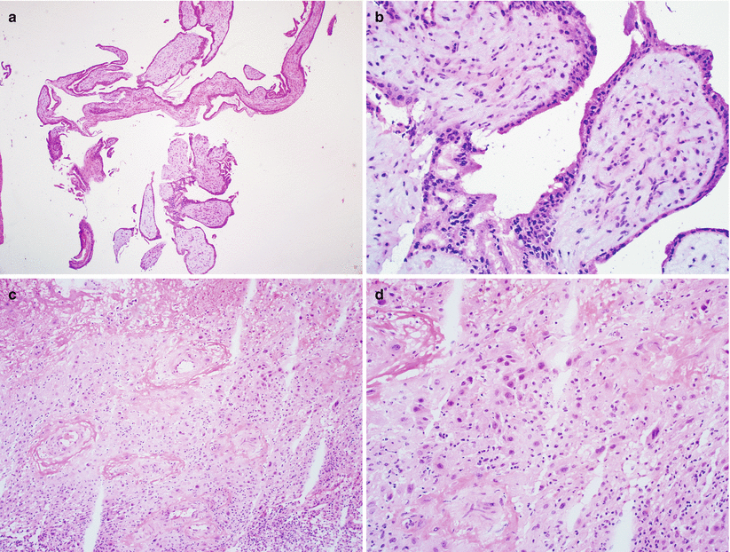
Fig. 6.1
Histological evidence of intrauterine pregnancy. Note the presence of chorionic villi (a, b) and implantation site intermediate trophoblast (c, d) in this uterine curettage specimen
Diagnostic pitfalls/key intraoperative consultation issues
Intrauterine pregnancy is confirmed by the presence of any of the three gestational tissue types: chorionic villi, fetal parts, or implantation site intermediate trophoblast.
Edematous endometrial and endocervical tissue fragments may be misinterpreted as chorionic villi; however, this pitfall can be avoided by recognition of cytotrophoblast and syncytiotrophoblast on the villous surface.
Presence of rare syncytiotrophoblast—without other gestational tissues—in an endometrial curettage does not rule out an ectopic pregnancy.
Hydatidiform Mole
Hydatidiform moles are nonneoplastic proliferations of the villous trophoblast, characterized by enlarged hydropic villi along with trophoblastic hyperplasia. They are incompatible with fetal survival and are genetically defined by their unique parental chromosome contributions. The two main subtypes are complete hydatidiform mole (CHM) and partial hydatidiform mole (PHM). Invasive hydatidiform mole is characterized by myometrial invasion. Persistent GTD is a clinical diagnosis and includes invasive mole and metastatic mole that may develop following either CHM or PHM.
Complete Hydatidiform Mole (CHM) [1, 2]
Clinical features
Patients with fully developed CHM present in the second trimester of pregnancy with vaginal bleeding, uterine enlargement, markedly elevated serum human chorionic gonadotropin (hCG), hyperemesis, toxemia, and hyperthyroidism.
Very early complete moles are frequently evacuated without clinical suspicion as missed abortion and without abnormally elevated serum hCG.
Gross pathology
Diffuse villous hydrops.
No fetal parts present.
Early evacuation of complete molar tissue before the second trimester typically lacks a gross lesion.
Microscopic features (Fig. 6.2)
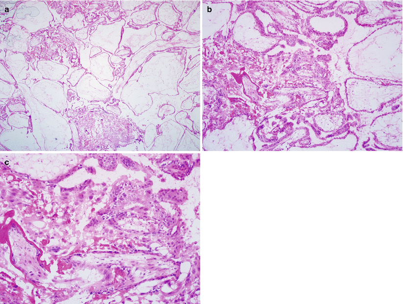
Fig. 6.2
Well-developed complete mole. Note the diffuse villous hydrops with cistern formation (a), marked, circumferential villous trophoblastic hyperplasia, (b) and nuclear atypia of the trophoblast (c)
Markedly edematous villi with central cistern formation.
Diffuse circumferential trophoblastic hyperplasia involving both cytotrophoblast and syncytiotrophoblast.
Presence of cytological atypia.
Absence of nucleated fetal red blood cells and fetal parts.
Early complete mole shows normal-sized chorionic villi with abnormal “cauliflower-like” or polypoid configurations. The villous stroma is characteristically hypercellular, composed of stellate fibroblasts embedded in a bluish myxoid matrix with prominent karyorrhexis. Trophoblastic hyperplasia may be focally present or absent.
Differential diagnosis
Partial mole
Hydropic non-molar abortion
Diagnostic pitfalls/key intraoperative consultation issues
Well-developed CHM presents with diffusely hydropic villi, which is not present in PHM and non-molar conditions.
Separation of very early complete mole from hydropic non-molar gestations, early gestational sac, and PHM is often challenging but not crucial at the time of intraoperative consultation.
Partial Hydatidiform Mole (PHM) [1, 3]
Clinical features
Majority of patients present in the late first trimester or early second trimester with missed or incomplete abortion.
Uterine size is usually small or appropriate for gestational age.
Serum hCG is usually moderately elevated and a fetus may be seen on ultrasound.
Gross pathology
Normal-sized villi admixed with edematous, enlarged ones
Frequent gestational sac or fetal parts
Stay updated, free articles. Join our Telegram channel

Full access? Get Clinical Tree


