2nd most common primary liver cancer after hepatocellular carcinoma
Has been increasing around world, including USA
Very prevalent in Asia, particularly in northeastern Thailand
Equal frequency in men and women
• Well-known risk factors include liver fluke infection, primary sclerosing cholangitis, hepatolithiasis, Thorotrast exposure, congenital anomalies of bile ducts
• Serum level of CA19-9 is commonly elevated
• Most patients are diagnosed with advanced stages of disease
Dismal prognosis
Microscopic
• Well- to moderately differentiated adenocarcinoma
Desmoplastic stroma
Frequently shows perineural invasion
Mucin typically present
CK19, CK7 positive
• Neoplastic cells can form glands, solid nests, cords, or papillary structures
Top Differential Diagnoses
• Hepatocellular carcinoma
• Metastatic adenocarcinoma
• Epithelioid hemangioendothelioma
• Bile ductular reaction or atypical biliary epithelium due to inflammation
• Benign hamartoma
• Biliary adenofibroma
• Hyperplasia of peribiliary glands
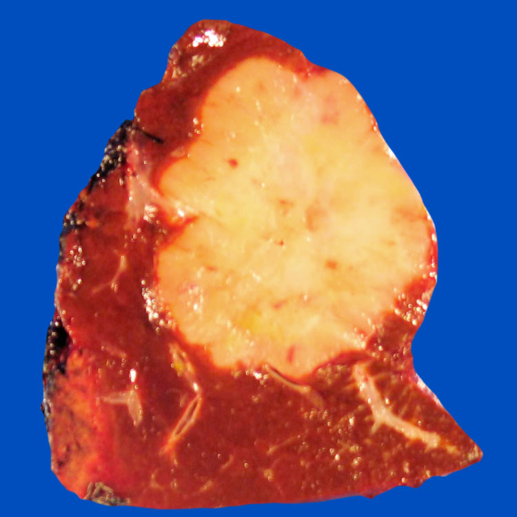
Intrahepatic Cholangiocarcinoma Arising in Noncirrhotic Liver Gross photograph shows a white-tan, firm, and distinct mass in a noncirrhotic liver.
Intrahepatic Cholangiocarcinoma in Cirrhosis A white, green to tan, irregular, firm mass is shown in this case of hepatitis C-associated cirrhosis. There is increased risk for cholangiocarcinoma in cirrhosis.
CECT of Intrahepatic Cholangiocarcinoma Axial reformatted CECT in portal venous phase shows a heterogeneous hepatic mass within a noncirrhotic liver. Satellite lesions of similar appearance are present as well.
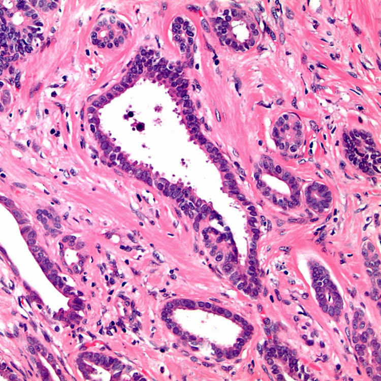
Well-Differentiated Appearance H&E shows infiltrative, well-formed glands with minimal nuclear atypia in a prominent fibrous stroma.
TERMINOLOGY
Definitions
• Primary adenocarcinoma arising from biliary epithelium
ETIOLOGY/PATHOGENESIS
Multistep Carcinogenesis
• Chronic inflammation may be common pathogenic pathway
• Wide array of genetic changes have been described, including TP53 and KRAS mutations
• Mutations in isocitrate dehydrogenase (IDH) 1 and 2
Observed in 25-30% of cases
– Uncommon in extrahepatic cases and adenocarcinomas of other gastrointestinal sites
CLINICAL ISSUES
Epidemiology
• Incidence
2nd most common primary liver cancer after hepatocellular carcinoma
Varies widely worldwide; more prevalent in East Asia than in Western countries
• Age
Average at presentation: 60 years
• Sex
Equal frequency in men and women
• Ethnicity
Very prevalent in Asia, particularly in Northeastern Thailand (associated with liver fluke infestation), East Asia
Presentation
• 10-20% of primary liver malignancies
Incidence and mortality rates have been increasing in several regions around world
Incidence has also increased 3x in past few decades in USA
• Most patients diagnosed with advanced stages of disease


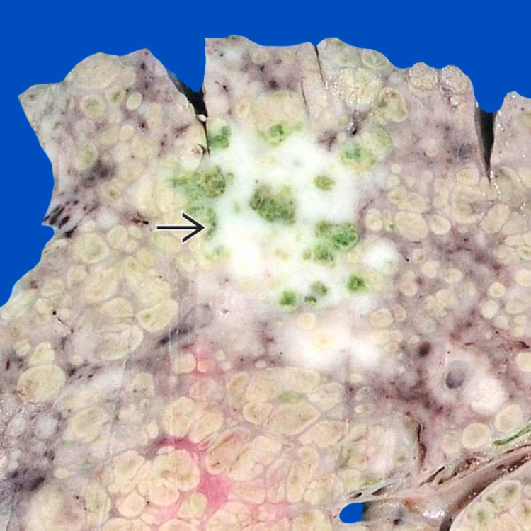
 is shown in this case of hepatitis C-associated cirrhosis. There is increased risk for cholangiocarcinoma in cirrhosis.
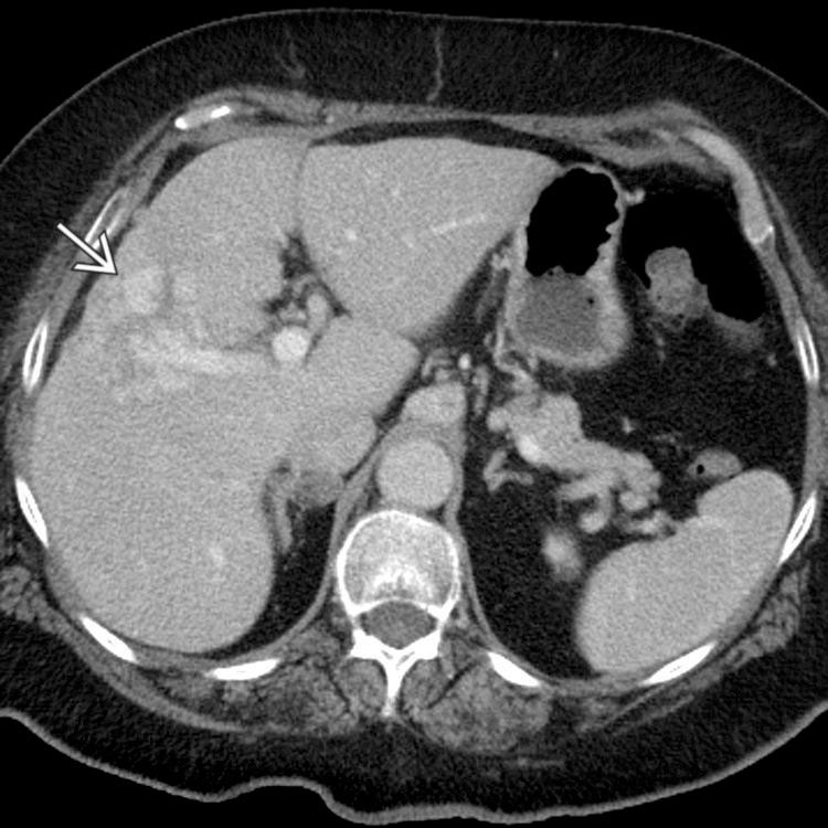
 within a noncirrhotic liver. Satellite lesions of similar appearance are present as well.