Intracholecystic Papillary-Tubular Neoplasms

 Includes neoplastic lesions previously classified as adenomas (both intestinal and pyloric types), papillary carcinoma in situ, papillomatosis


Clinical Issues


• Many patients asymptomatic, and ICPN discovered incidentally at cholecystectomy

• Associated with invasive carcinoma in 50% of cases at diagnosis

• 5-year survival of 60% for cases with associated invasive carcinoma, 78% for noninvasive lesions


Macroscopic


• Most cases are solitary; 1/3 multiple

• May become detached from lumen and grossly mimic gallstones or biliary sludge

• Average size: ∼ 2 cm; reported as large as 7 cm


Microscopic


• Classified as tubular (> 75%), papillary (> 75%), or tubulopapillary (25-75% each pattern)
• Cell types: Gastric (pyloric, foveolar), intestinal, biliary, oncocytic

image Pyloric type has lowest association with high-grade dysplasia and carcinoma (∼ 15%)

image Intestinal type resembles colonic adenomas

image Biliary type accounts for ∼ 50% of ICPN, frequently associated with high-grade dysplasia and carcinoma


Diagnostic Checklist


• Entire lesion should be submitted for microscopic examination to rule out associated invasive carcinoma

image
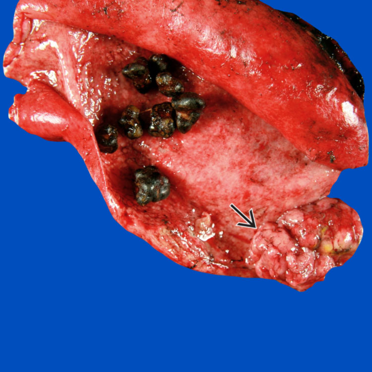
Gallbladder ICPN
An exophytic, lobulated intraluminal polypoid intracholecystic papillary neoplasm image is present in this cholecystectomy specimen, associated with chronic cholecystitis and gallstones.


image
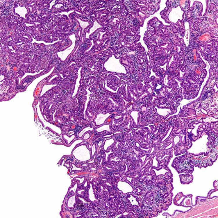
Pyloric Type
This large pyloric-type ICPN forms a polypoid lesion in the lumen of the gallbladder. It is composed of tightly packed pyloric-type glands and tubules.

image
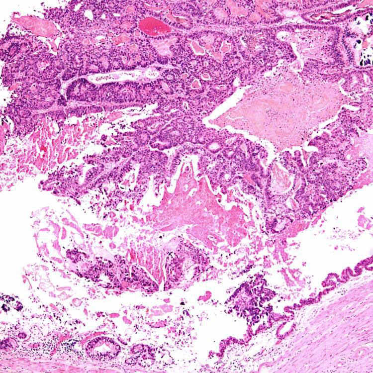
Detached ICPN
Pedunculated ICPNs often become detached, as shown here, and may resemble biliary sludge or soft stones in the lumen of the gallbladder. This intestinal-type ICPN had high-grade dysplasia.

image
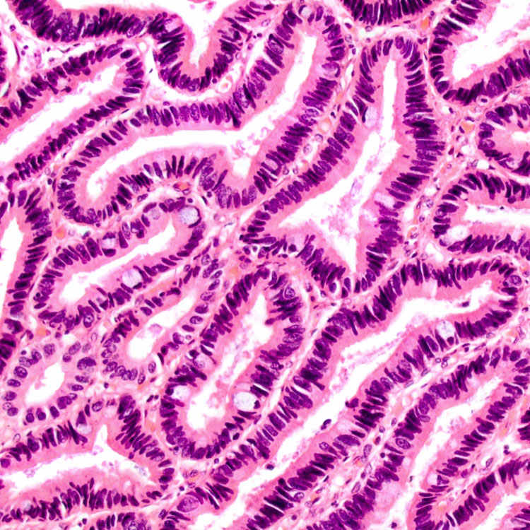
Intestinal Type
This intestinal-type ICPN shows low-grade dysplasia, similar to a colonic adenoma. Scattered goblet cells are present.


TERMINOLOGY


Abbreviations


• Intracholecystic papillary-tubular neoplasms (ICPN)


Synonyms


• Tumoral intraepithelial neoplasms


Definitions


• Exophytic or polypoid neoplastic epithelial proliferation in gallbladder
image Includes neoplastic lesions previously classified as adenomas (both intestinal and pyloric types), papillary carcinoma in situ, papillomatosis

image ≥ 1 cm

– “Incipient ICPN” has been suggested for adenomas that have dysplasia but are < 1 cm

image Biliary, foveolar, pyloric, intestinal, and oncocytic cell types

image Noninvasive by definition

image Distinct from adjacent mucosa


CLINICAL ISSUES


Epidemiology


• Age
image Mean: 61 years

• Sex
image Female predominance (F:M = 2:1)

• Ethnicity
image More common in Asia


Presentation


• Often discovered incidentally at cholecystectomy
• When symptomatic, patients typically present with right upper quadrant pain

image Jaundice, biliary obstruction can occur with multiple lesions or location near neck of gallbladder

• Only ∼ 20% of cases associated with gallstones

• ∼ 20% of patients have other neoplasms at time of diagnosis
image Most commonly GI tract and pancreatic tumors


Treatment


• Surgical approaches
image Cholecystectomy is curative in most cases with noninvasive lesions

image New primary lesions may subsequently develop in biliary tree


Prognosis


• Invasive carcinoma present in > 50% of cases at diagnosis
image High-risk features

– Extent of high-grade dysplasia

– Extent of papillary component

– Size of lesion

– Intestinal or pancreatobiliary phenotype

image Most commonly pancreaticobiliary type adenocarcinomas

image 5-year survival 60% in ICPN with invasive component

image Only minority of gallbladder adenocarcinomas associated with precursor ICPN, however

Only gold members can continue reading. Log In or Register to continue

Stay updated, free articles. Join our Telegram channel

Apr 20, 2017 | Posted by in PATHOLOGY & LABORATORY MEDICINE | Comments Off on Intracholecystic Papillary-Tubular Neoplasms

Full access? Get Clinical Tree

Get Clinical Tree app for offline access