Patient Story
A parent brings in a 4-year-old boy suffering with anal itching. On examination the physician finds several excoriations around the anus and suspects pinworms. The physician then applies scotch tape to the perianal area and places the tape on a glass slide. Review of the slide demonstrates adult worms and ova of Enterobius vermicularis (pinworms) (Figure 214-1). The boy is treated with a single dose of chewable mebendazole and his symptoms resolve. The parent is told to repeat the mebendazole dose in 2 weeks to increase the long-term cure rate. If the scotch tape test were negative, the physician could choose to treat empirically as mebendazole is a very safe medication. Another option is to test again having the parent apply the scotch tape to the boy’s perianal area first thing in the morning and bring that back to the office (the yield is higher in the morning).
Introduction
Intestinal parasites are most common in places with warmer temperatures and high humidity, poor sanitation and unclean water, and a large number of individuals (especially children) living in close proximity. In general, the parasites are either asymptomatic or cause symptoms related to their presence in the GI tract. Several migrate through the lungs and can also cause pulmonary symptoms during the migration. Diagnoses are made by history of worms being seen by the patient or parents or by laboratory examination for ova and parasites in the stool.
Epidemiology
- Nematoda is the phylum that contains pinworms, hookworms, Ascaris, Strongyloides, and whipworms.
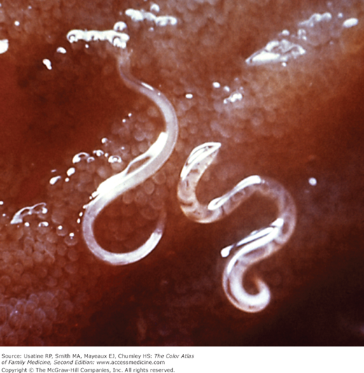
- E. vermicularis (pinworm) is the most prevalent nematode in the United States. Populations at risk include preschool and school aged children, institutionalized persons, and household members of persons with pinworm infection1 (Figure 214-1).
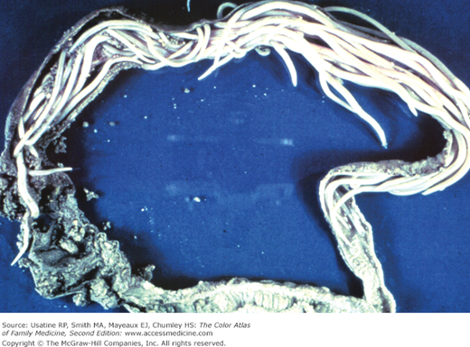
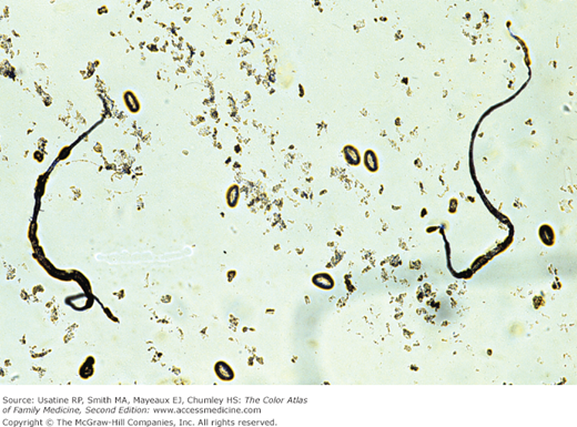
- Necator americanus (hookworm) is found predominately in the Americas and Australia, and is the second most common nematode identified in stool studies in the United States1 (Figures 214-2 and 214-3). Ancylostoma duodenale (hookworm) is found mostly in southern Europe, North Africa, the Middle East, and Asia.1
- Ascaris lumbricoides is the largest and most common roundworm found in humans in the world; although less common in the United States, it is seen mostly in the rural southeast. It is found in tropical and subtropical areas, including the southeastern rural United States (Figures 214-4 and 214-5).1
- Strongyloides stercoralis is seen mostly in tropical and subtropical areas, but can be found in temperate areas, including the southern United States (Figure 214-6). It is more frequently found in rural areas, institutional settings, and lower socioeconomic groups.1
- Trichuris trichiura (whipworm) is the third most common roundworm found in humans worldwide. Infections are more frequent in areas with tropical weather and poor sanitation practices, and among children (Figure 214-7). It is estimated that 800 million people are infected worldwide. Trichuriasis occurs in the southern United States.1
- E. vermicularis (pinworm) is the most prevalent nematode in the United States. Populations at risk include preschool and school aged children, institutionalized persons, and household members of persons with pinworm infection1 (Figure 214-1).
- Cestodes (tapeworm) are a class in the phylum Platyhelminthes that contains Taenia solium (pork tapeworm).
- T. solium is found worldwide where pigs and humans live in close proximity.
- Protozoa is the kingdom of one-celled organisms that includes Giardia lamblia and Entamoeba histolytica.
- G. lamblia (Giardia intestinalis) is the most common parasite infection worldwide and the second most common in the United States (after pinworm), causing 2.5 million infections annually (Figure 214-8).1
- E. histolytica is seen worldwide, with higher incidence in developing countries. In the United States, risk groups include men who have sex with men, travelers and recent immigrants, and institutionalized populations.1
- G. lamblia (Giardia intestinalis) is the most common parasite infection worldwide and the second most common in the United States (after pinworm), causing 2.5 million infections annually (Figure 214-8).1